. 2021 Jun:273:120810.
doi: 10.1016/j.biomaterials.2021.120810. Epub 2021 Apr 9.

miR-145 micelles mitigate atherosclerosis by modulating vascular smooth muscle cell phenotype

Affiliations

miR-145 micelles mitigate atherosclerosis by modulating vascular smooth muscle cell phenotype

Deborah D Chin et al. Biomaterials. 2021 Jun.

Abstract

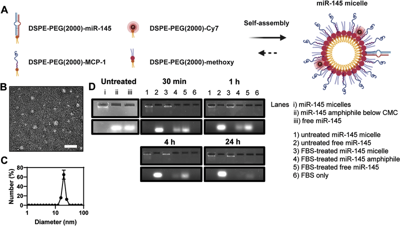
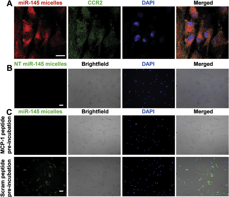
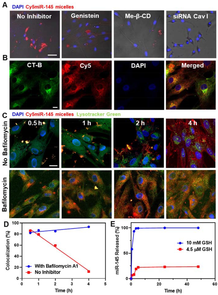
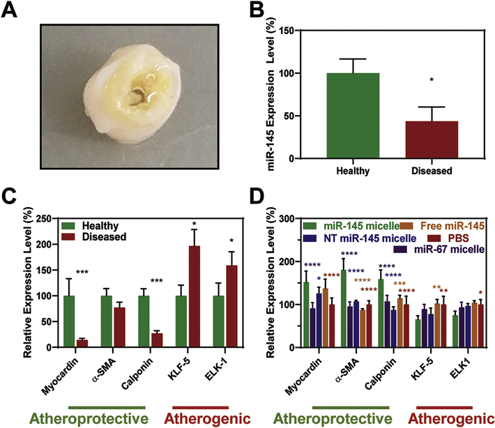
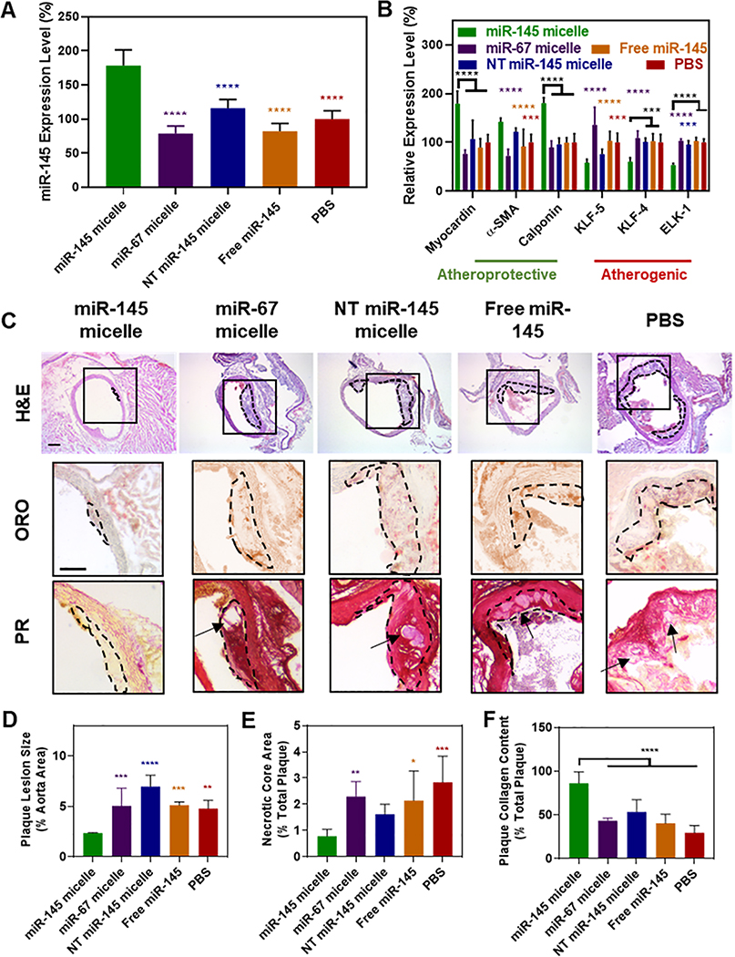
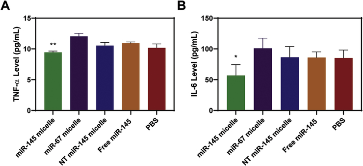
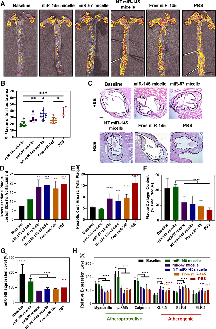
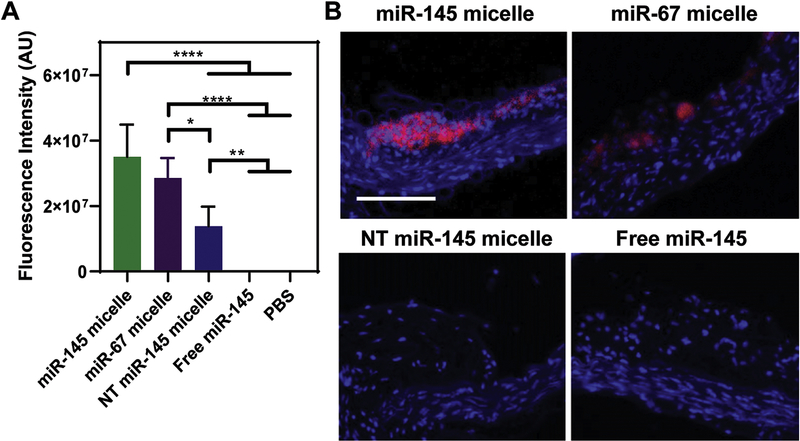
In atherosclerosis, resident vascular smooth muscle cells (VSMCs) in the blood vessels become highly plastic and undergo phenotypic switching from the quiescent, contractile phenotype to the migratory and proliferative, synthetic phenotype. Additionally, recent VSMC lineage-tracing mouse models of atherosclerosis have found that VSMCs transdifferentiate into macrophage-like and osteochondrogenic cells and make up to 70% of cells found in atherosclerotic plaques. Given VSMC phenotypic switching is regulated by microRNA-145 (miR-145), we hypothesized that nanoparticle-mediated delivery of miR-145 to VSMCs has the potential to mitigate atherosclerosis development by inhibiting plaque-propagating cell types derived from VSMCs. To test our hypothesis, we synthesized miR-145 micelles targeting the C-C chemokine receptor-2 (CCR2), which is highly expressed on synthetic VSMCs. When miR-145 micelles were incubated with human aortic VSMCs in vitro, >90% miR-145 micelles escaped the lysosomal pathway in 4 hours and released the miR cargo under cytosolic levels of glutathione, an endogenous reducing agent. As such, miR-145 micelles rescued atheroprotective contractile markers, myocardin, α-SMA, and calponin, in synthetic VSMCs in vitro. In early-stage atherosclerotic ApoE-/- mice, one dose of miR-145 micelles prevented lesion growth by 49% and sustained an increased level of miR-145 expression after 2 weeks post-treatment. Additionally, miR-145 micelles inhibited 35% and 43% plaque growth compared to free miR-145 and PBS, respectively, in mid-stage atherosclerotic ApoE-/- mice. Collectively, we present a novel therapeutic strategy and cell target for atherosclerosis, and present miR-145 micelles as a viable nanotherapeutic that can intervene atherosclerosis progression at both early and later stages of disease.

Keywords: Atherosclerosis; Micelle; Nanomedicine; Peptide; Smooth muscle cell; microRNA.


Conflict of interest statement

Competing interests

There are no competing interests to state.

Declaration of interests

The authors declare that they have no known competing financial interests or personal relationships that could have appeared to influence the work reported in this paper.

Figures

Fig. 1.
Fig. 1.. Schematic and characterization of miR-145 micelles.
(A) Schematic of the self-assembly and multifunctionality of miR-145 micelles. (B) TEM images of miR-145 micelles. Scale bar = 200 nm. (C) DLS of miR-145 micelles. (D) Gel shift assays confirm miR-145 is protected when incorporated into micelles, nuclease degradation for up to 24 hours.
Fig. 2.
Fig. 2.
miR-145 micelle binding on VSMCs. (A) miR-145 micelles bind to CCR2 on VSMCs. Scale bar = 25 μm. (B) NT miR-145 micelles show no binding to diseased patient VSMCs. Scale bar = 100 μm. (C) Preincubation with MCP-1 significantly reduces miR-145 micelle binding, whereas preincubation with scrambled MCP-1 peptide does not affect miR-145 micelle binding to VSMCs. Scale bar = 100 μm.
Fig. 3.
Fig. 3.
Cellular internalization, endosomal escape, and release of miR-145 micelles. (A) Fluorescence images of hASMCs incubated with Cy5miR-145 micelles (red) and endocytosis inhibitors. Scale bar = 50 μm. (B) hASMCs incubated with miR-145 micelles and treated with CT-B confirm micelle internalization is through caveolae-mediated endocytosis. Channels: Cy5-micelles (red) and CT-B (green). Scale bar = 20 μm. Time-dependent endosomal escape of Cy5miR-145 micelle in hASMCs (C) without and with bafilomycin A1. Nuclei were stained with DAPI (blue). Scale bar = 20 μm. (D) Percent colocalization of miR and endosomes. (E) Release profiles of miR-145 from micelles show release of miR-145 at intracellular GSH concentrations (10 mM).
Fig. 4.
Fig. 4.
miR-145 micelles rescue VSMC contractile markers and silence synthetic markers. (A) Patient-derived, tibial artery samples with significant plaque build-up used to isolate VSMCs. These VSMCs have (B) lower miR-145 expression compared to VSMCs from healthy tissue (N = 3; *p < 0.05) and (C) lower endogenous expression of contractile markers and higher expression of synthetic markers (N = 4; *p < 0.05, ***p < 0.001 compared to healthy samples). (D) miR-145 micelle treatment on VSMCs derived from diseased arteries rescue contractile markers and downregulate synthetic markers (N = 4; *p < 0.05, ***p < 0.001, relative to miR-145 micelles).
Fig. 5.
Fig. 5.
miR-145 micelles promote contractile VSMCs and prevent atherogenesis in early-stage disease. (A) miR-145 and (B) myocardin, α-SMA, calponin, KLF-5, KLF-4, and ELK-1 expression in VSMCs derived from early-stage atherosclerotic mice (N = 6). (C) Representative histological cross-sections of the ascending aorta stained with H&E, ORO, and PR (plaques: dotted black line, necrotic core: arrows). Scale bar = 200 μm. miR-145 micelle treatment decreases (D) plaque lesion size and (E) necrotic core area, while preserving (F) collagen content in plaques (Note: N = 6 for all treatment groups, except miR-145 micelles with N = 4 as two mice did not form plaques, *p < 0.05, **p < 0.01, ***p < 0.001, ****p < 0.0001 compared to miR-145 micelles).
Fig. 6.
Fig. 6.
Anti-inflammatory effects of miR-145 micelles. Mice treated with miR-145 micelles show a decrease in serum (A) TNF-α and (B) IL-6 in early-stage ApoE−/− mice (N = 6; *p < 0.05, **p < 0.01).
Fig. 7.
Fig. 7.. miR-145 micelles promote contractile VSMCs and reduce plaque progression in mid-stage atherosclerotic mice.
(A) En face ORO stains of arteries show fewer plaques (yellow outline) in miR-145 micelle-treated mice and the baseline control (N > 5). (B) Quantification of ORO en face staining (N > 5). (C) Representative histological images of the aortic root stained with H&E showing plaques (dotted black line). Scale bar = 200 μm. Analysis of (D) cross-sectional plaque lesion size, (E) necrotic core area, and (F) collagen (N > 11 for treatment groups, N = 6 for baseline). (G) miR-145 expression and (H) mRNA expression of contractile and synthetic phenotype markers in VSMCs from mid-stage atherosclerotic mice (N = 6; *p < 0.05, **p < 0.01, ***p < 0.001, ****p < 0.0001 compared to miR-145 micelles).
Fig. 8.
Fig. 8.
In vivo targeting of miR-145 micelles. (A) Fluorescence quantification in aortas of atherosclerotic mice show miR-145 micelles accumulate to the greatest degree compared to miR-67 micelles and NT miR-145 micelles (N = 6). (B) Histological analyses show miR-145 micelles (red) accumulate in plaques. Scale bar = 100 μm.
Fig. 9.
Fig. 9.. In vivo biocompatibility of miR-145 micelles.
(A) H&E staining of major organs show no significant morphological changes associated with miR-145 micelles in mid-stage atherosclerotic mice (NT micelle = NT miR-145 micelle). Scale bar = 100 μm. (B) No significant change in body weight was observed for all groups, demonstrating the safety and tolerance of micelles (N = 12). (C) BUN and creatinine measurements confirm kidney health of mice and safety of micelles (N = 6).

References

    1. Brozovich FV, et al., Mechanisms of Vascular Smooth Muscle Contraction and the Basis for Pharmacologic Treatment of Smooth Muscle Disorders. Pharmacol Rev, 2016. 68(2): p. 476–532. - PMC - PubMed
    1. Bennett MR, Sinha S, and Owens GK, Vascular Smooth Muscle Cells in Atherosclerosis. Circ Res, 2016. 118(4): p. 692–702. - PMC - PubMed
    1. Gomez D and Owens GK, Smooth muscle cell phenotypic switching in atherosclerosis. Cardiovascular research, 2012. 95(2): p. 156–164. - PMC - PubMed
    1. Shankman LS, et al., KLF4-dependent phenotypic modulation of smooth muscle cells has a key role in atherosclerotic plaque pathogenesis. Nature Medicine, 2015. 21: p. 628. - PMC - PubMed
    1. Basatemur GL, et al., Vascular smooth muscle cells in atherosclerosis. Nature Reviews Cardiology, 2019. 16(12): p. 727–744. - PubMed

Publication types