Review
. 2021 Apr 23;19(1):47.
doi: 10.1186/s12964-021-00730-1.

The exosome journey: from biogenesis to uptake and intracellular signalling

Affiliations
Review

The exosome journey: from biogenesis to uptake and intracellular signalling

Sonam Gurung et al. Cell Commun Signal. .

Abstract

The use of exosomes in clinical settings is progressively becoming a reality, as clinical trials testing exosomes for diagnostic and therapeutic applications are generating remarkable interest from the scientific community and investors. Exosomes are small extracellular vesicles secreted by all cell types playing intercellular communication roles in health and disease by transferring cellular cargoes such as functional proteins, metabolites and nucleic acids to recipient cells. An in-depth understanding of exosome biology is therefore essential to ensure clinical development of exosome based investigational therapeutic products. Here we summarise the most up-to-date knowkedge about the complex biological journey of exosomes from biogenesis and secretion, transport and uptake to their intracellular signalling. We delineate the major pathways and molecular players that influence each step of exosome physiology, highlighting the routes of interest, which will be of benefit to exosome manipulation and engineering. We highlight the main controversies in the field of exosome research: their adequate definition, characterisation and biogenesis at plasma membrane. We also delineate the most common identified pitfalls affecting exosome research and development. Unravelling exosome physiology is key to their ultimate progression towards clinical applications. Video Abstract.

Keywords: Endocytosis; Exosomes; Extracellular vesicles; Intercellular communication; Lipid rafts; Multivesicular bodies; Rab GTPases; Targeting; Tetraspanins.


Conflict of interest statement

The authors declare that they have no competing interests.

Figures
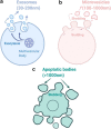
Fig. 1
Fig. 1
Extracellular vesicles (EVs) classification. The three different classes of EVs are depicted. a Exosomes are generated through the endocytic pathway and are released via exocytosis, are spherical in shape and have size range of 30–200 nm of diameter. b Microvesicles (MVs) are released through budding from plasma membrane, are irregular in shape and range in size between 100–1000 nm of diameter. c Apoptotic bodies are released through blebbing by cells undergoing apoptosis and are > 1000 nm in size
Fig. 2
Fig. 2
Composition of exosomes. Exosomes are composed of various proteins: transmembrane proteins such as tetraspanins, antigen presenting molecules, glycoproteins and adhesion molecules; proteins in exosome lumen such as heat shock proteins (Hsp), cytoskeletal proteins, ESCRT components, membrane transport, fusion proteins, growth factors and cytokines. Exosomes also comprise of multiple lipids such as cholesterol, ceramides, sphingomyelin, phosphatidylinostol (PI), phosphatidylserine (PS), phosphatidylcholine (PC), phosphatidylethanolamine (PE) and gangliosides (GM) along with nucleic acids such as mRNA, miRNA, non-coding RNA and DNA in their lumen. Hsc = Heat shock cognate; TSG = tumor suspectibility gene; TNF = tumor necrosis factor; TGF = Transforming growth factor; TRAIL = TNF-related apoptosis-inducing ligand; FasL = Fas ligand; TfR = Transferrin receptor
Fig. 3
Fig. 3
Exosome biogenesis. Within the endosomal system, [1] internalised cargoes are [2] sorted into early endosomes, [3] which then mature into late endosomes or multivesicular bodies. Late endosomes/multivesicular bodies are specialised endosomal compartments rich in intraluminal vesicles (ILVs), which sequester proteins, lipids, and cytosolic compartments and potential exosome cargoes. [4] Cargoes are also delivered from trans-Golgi network and possibly from cytosol. [5] Multivesicular bodies containing exosome cargoes get [5] transported to the plasma membrane, [6] fuse with the cell surface and [7] the ILVs then get secreted as exosomes. ER: Endoplasmic Reticulum; MVB: Multivesicular Bodies; PM: Plasma membrane
Fig. 4
Fig. 4
Exosome signalling by direct interaction or membrane fusion. Upon reaching the target cells, a membrane receptors within the exosome surface and plasma membrane of target cells can interact inducing downstream signalling cascade in the recipient cell. b Exosomes membrane can also fuse with the plasma membrane and release their contents into the cytosol directly
Fig. 5
Fig. 5
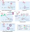
Exosome internalisation. Exosomes are internalised by the recipient cells and fuse with the intracellular compartments/endosomal pathway for cargo release. Exosomes can be internalised by a clathrin-mediated endocytosis, b lipid-raft mediated, c caveolin-mediated endocytosis, d phagocytosis or e micropinocytosis. These pathways are not always mutually exclusive and can co-exist for the internalisation of a same set of exosomes
Fig. 6
Fig. 6
Exosome biology. [1] Exosomes are generated through the formation of ILVs in the late endocytic pathway and [2] gets secreted via exocytosis from the plasma membrane. Upon reaching the target recipient cell, [3] exosomes either interact with the surface molecules of recipient cell to induce downstream signalling or [4] fuse with the plasma membrane to release their contents into cytosol or [5] get internalised via various routes. [6] Upon internalisation, exosomes are addressed in the early endosome, then late endosomes or MVBs and undergo multiple fates. [7] The exosome contents can get released into the nucleus or ER, [8] leak into cytosol or [9] get degraded in the lysosomes. [10] Another possibility include release back to the extracellular space through the recycling endosome. ILV: Intraluminal Vesicles; EE: Early Endosomes, RE: Recycling endosomes, MVB: Multivesicular Bodies, ER: Endoplasmic Reticulum

References

    1. Deatherage BL, Cookson BT. Membrane vesicle release in bacteria, eukaryotes, and archaea: a conserved yet underappreciated aspect of microbial life. Infect Immun. 2012;80(6):1948–1957. doi: 10.1128/IAI.06014-11. - DOI - PMC - PubMed
    1. Doyle LM, Wang MZ. Overview of extracellular vesicles, their origin, composition, purpose, and methods for exosome isolation and analysis. Cells. 2019;8(7):727. doi: 10.3390/cells8070727. - DOI - PMC - PubMed
    1. Johnstone RM, Adam M, Hammond JR, Orr L, Turbide C. Vesicle formation during reticulocyte maturation. Association of plasma membrane activities with released vesicles (exosomes) J Biol Chem. 1987;262(19):9412–9420. doi: 10.1016/S0021-9258(18)48095-7. - DOI - PubMed
    1. Valadi H, Ekström K, Bossios A, Sjöstrand M, Lee JJ, Lötvall JO. Exosome-mediated transfer of mRNAs and microRNAs is a novel mechanism of genetic exchange between cells. Nat Cell Biol. 2007;9(6):654–659. doi: 10.1038/ncb1596. - DOI - PubMed
    1. Pegtel DM, Gould SJ. Exosomes. Annu Rev Biochem. 2019;88:487–514. doi: 10.1146/annurev-biochem-013118-111902. - DOI - PubMed

Publication types

LinkOut - more resources