Review
. 2021 Sep;37(9):790-802.
doi: 10.1016/j.pt.2021.03.005. Epub 2021 Apr 20.

Single-cell deconstruction of stem-cell-driven schistosome development

Affiliations
Review

Single-cell deconstruction of stem-cell-driven schistosome development

Dania Nanes Sarfati et al. Trends Parasitol. 2021 Sep.

Abstract

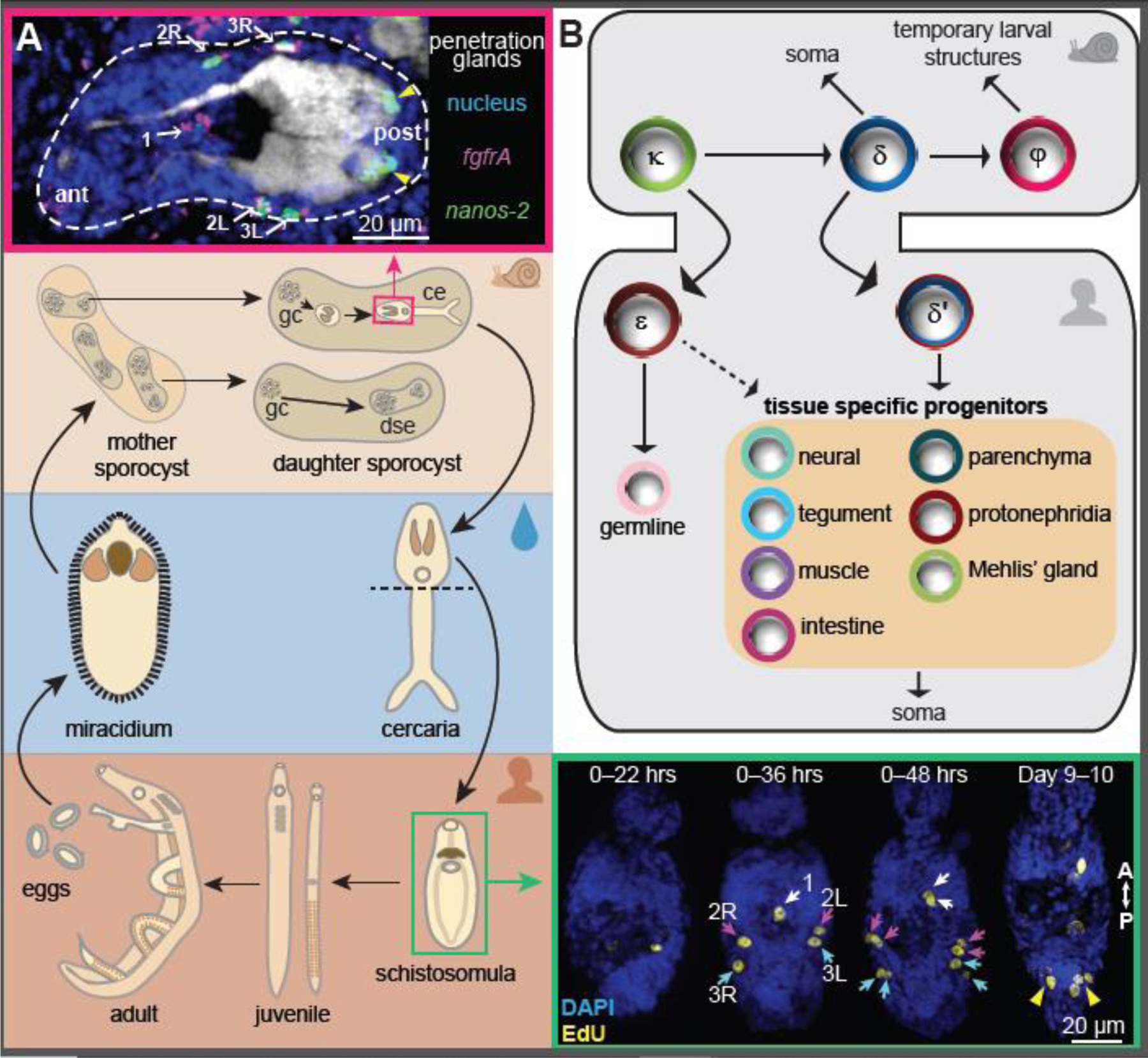
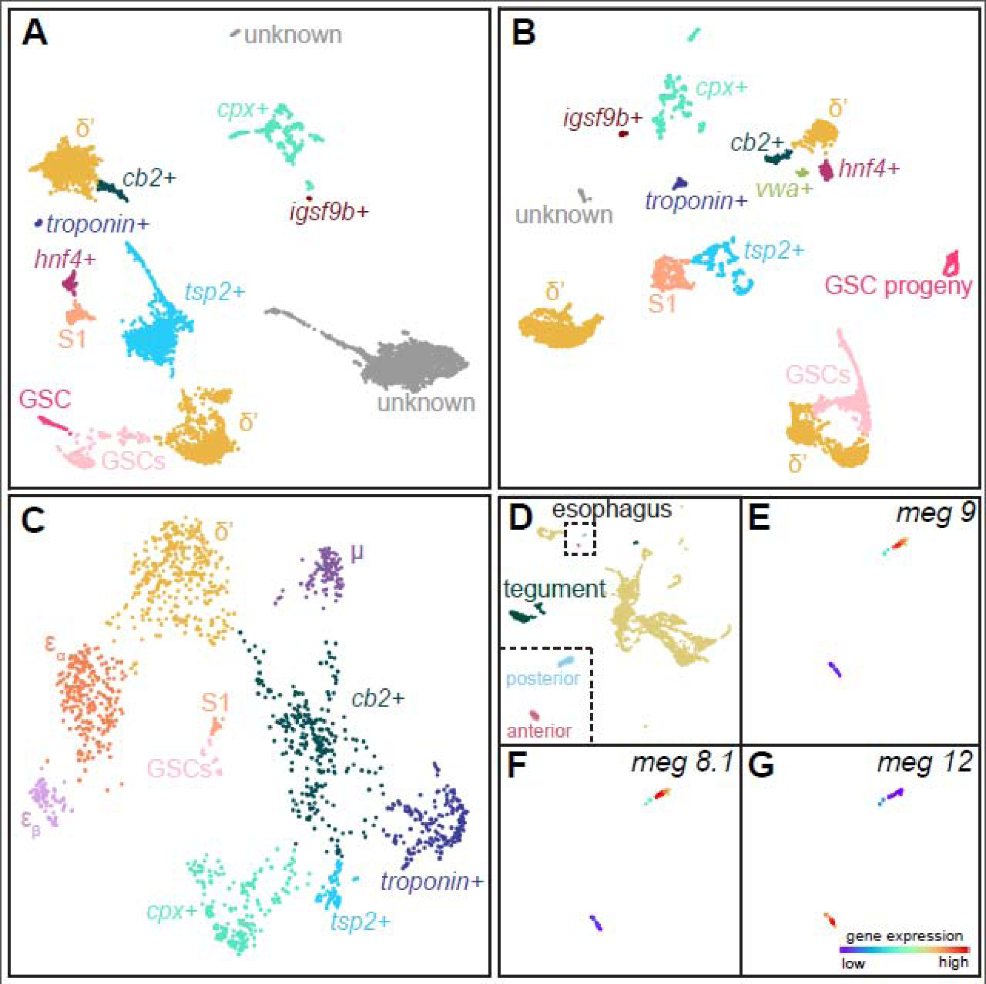
Schistosomes cause one of the most devastating neglected tropical diseases, schistosomiasis. Their transmission is accomplished through a complex life cycle with two obligate hosts and requires multiple radically different body plans specialized for infecting and reproducing in each host. Recent single-cell transcriptomic studies on several schistosome body plans provide a comprehensive map of their cell types, which include stem cells and their differentiated progeny along an intricate developmental hierarchy. This progress not only extends our understanding of the basic biology of the schistosome life cycle but can also inform new therapeutic and preventive strategies against the disease, as blocking the development of specific cell types through genetic manipulations has shown promise in inhibiting parasite survival, growth, and reproduction.

Keywords: cell type atlas; development; life cycle; schistosome; single-cell sequencing; stem cells.


Conflict of interest statement

Declaration of interests The authors declare no competing interests.

Figures

Figure 1.
Figure 1.. Schistosomes remodel their body plans multiple times throughout their life cycle.
(A) Schematics showing the schistosome life cycle as detail in Box 1. dse.: daughter sporocyst embryo; gc: germinal cells; ce: cercarial embryo. Insets: microscopy images showing that germinal cells are transmitted through the cercarial heads to initiate the intramammalian development. Images correspond to boxed stages in the schematics, and are adapted from ref. . Top: a small number of germinal cells are packed at specific locations around the cercarial penetration glands. Arrows: germinal cells that are double positive for fgrfA and nanos-2 (δ-cells); arrowheads: nanos-2+ cells (κ-cells). Bottom right: these cells re-enter the cell cycle and proliferate in schistosomula to expand the stem cell pool. The five anterior stem cells (arrows) first enter the cell cycle and divide synchronously, whereas the two posterior stem cell clusters (arrowheads) begin to proliferate at a later time point. EdU labels newly synthesized DNA. (B) A model for the developmental hierarchy of schistosome germinal/stem cells across life cycle stages, which is adapted from ref. . Population specific genes are listed in Table 2.
Figure 2.
Figure 2.. Stem cell and esophageal subpopulations at the intramammalian stages.
Subclustering of ago2–1+ cells from adult male [22] (A) and virgin female [22] (B) parasites. Panel (C) shows juvenile ago2–1+ cells, adapted from ref. . Subpopulations of stem cells are annotated using gene expression signatures summarized in Table 2, and labeled by colors. We detected S1 cells in the adult male dataset, which are likely experimental contaminations, as S1-specific genes were shown to be absent in males [67]. (D) Analysis of the schistosomulum cell atlas [19] identifies a cluster containing the two tegumental progeny populations and fully differentiated tegumental cells (Table 3), and two distinct populations of esophageal cells corresponding to the anterior cell mass and posterior gland cells, respectively. Inset: a magnified view of the two esophageal clusters. Similar to many other known gland marker genes [49], the expression of posterior gland markers, meg-9 (E) and meg-8.1 (F), and anterior cell mass marker, meg-12 (G), is highly specific to each cluster. The manifold reconstruction was performed using Self-Assembling Manifolds (SAM) [20]. The SAM algorithm (version 0.7.5) was run using parameters ‘weight_PCs = False’ and ‘preprocessing = StandardScaler’. The SAM source code, tutorials, documentation, stem cell gene expression data, and subpopulation annotations are available through Github (https://github.com/atarashansky/self-assembling-manifold). While ago2–1+ cells form a distinct cluster in juveniles [21], they are distributed in multiple clusters in adults and separated based on their detected ago2–1 expression (>1 unique transcripts). Schistosomulum ago2–1+ cells lack elaborate subpopulation structures, and therefore are not shown.

References

    1. Hoffmann KF et al. (2014) Halting harmful helminthes: vaccines and new drugs are needed to combat parasitic worm infections. Science 346, 168–169 - PubMed
    1. Hotez PJ and Fenwick A (2009) Schistosomiasis in Africa: an emerging tragedy in our new global health decade. PLoS Negl. Trop. Dis 3, e485. - PMC - PubMed
    1. Bergquist R et al. (2017) Controlling schistosomiasis with praziquantel: how much longer without a viable alternative? Infect Dis Poverty 6, 74. - PMC - PubMed
    1. Doenhoff MJ et al. (2009) Praziquantel: Its use in control of schistosomiasis in sub-Saharan Africa and current research needs. Parasitology 136, 1825–1835 - PubMed
    1. Chan JD et al. (2017) The anthelmintic praziquantel is a human serotoninergic G-protein-coupled receptor ligand. Nat. Commun 8, 1910. - PMC - PubMed

Publication types