Review
. 2021 May-Jun;18(3):167-196.
doi: 10.21873/cgp.20251.

Interstitial Deletions Generating Fusion Genes

Affiliations
Review

Interstitial Deletions Generating Fusion Genes

Ioannis Panagopoulos et al. Cancer Genomics Proteomics. 2021 May-Jun.

Abstract

A fusion gene is the physical juxtaposition of two different genes resulting in a structure consisting of the head of one gene and the tail of the other. Gene fusion is often a primary neoplasia-inducing event in leukemias, lymphomas, solid malignancies as well as benign tumors. Knowledge about fusion genes is crucial not only for our understanding of tumorigenesis, but also for the diagnosis, prognostication, and treatment of cancer. Balanced chromosomal rearrangements, in particular translocations and inversions, are the most frequent genetic events leading to the generation of fusion genes. In the present review, we summarize the existing knowledge on chromosome deletions as a mechanism for fusion gene formation. Such deletions are mostly submicroscopic and, hence, not detected by cytogenetic analyses but by array comparative genome hybridization (aCGH) and/or high throughput sequencing (HTS). They are found across the genome in a variety of neoplasias. As tumors are increasingly analyzed using aCGH and HTS, it is likely that more interstitial deletions giving rise to fusion genes will be found, significantly impacting our understanding and treatment of cancer.

Keywords: Interstitial deletion; chromosome; cytogenetics; fusion gene; review.


Conflict of interest statement

The Authors declare that they have no potential conflicts of interest with regards to this study.

Figures
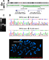
Figure 1
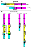
Figure 1. Formation of a fusion gene (Gene A-B) by an interstitial deletion and a chromosome translocation. (A) The deletion starts within Gene A and finishes within Gene B. Both genes are transcribed from centromere to telomere. The juxtaposition of the two genes by removal of the chromosome segment (yellow region) between them results in the chimeric Gene A-B consisting of the head (5’-end) of Gene A and the tail (3’-end) of Gene B. Loss of gene loci mapping in the yellow region, between the fusion partners, accompanies the fusion gene formation. (B). Formation of Gene A-B fusion by chromosome translocation between the two homologous chromosomes ChrZ-1 and ChrZ-2. Gene A-B is formed on der(ChrZ-1) whereas the reciprocal Gene B-A is formed on the der(ChrZ-2) chromosome. Duplication of gene loci mapped in the yellow region accompanies the reciprocal Gene B-A formation
Figure 2
Figure 2. Identification of a TRPS1-PLAG1 fusion gene which is generated by the interstitial deletion del(8)(q12q23). (A) Partial karyotype showing the del(8)(q12q23) and the normal chromosome 8 (breakpoints are shown by arrows). (B) Array comparative genomic hybridization showing the deletion in the q arm of chromosome 8. Based on the hg19 assembly, the deletion started at position Chr8:57120365 in intron1 of PLAG1 and ended at Chr8:116661489 in exon 1 of TRPS1. (C) Gel electrophoresis showing the amplified TRPS1-PLAG1 cDNA fragments. (D) Partial sequence chromatograms of the cDNA amplified fragment showing the junction positions of exon 1 of TRPS1 with exon 2 of PLAG1 and exon 1 of TRPS1 with exon 3 of PLAG1. E) FISH analysis on metaphase spreads with PLAG1 probe (red signal) and TRPS1 probe (green signal) showing that the TRPS1-PLAG1 fusion gene is on the del(8)(q12q23) (yellow signal). One copy of PLAG1 (red signal) and one of TRPS1 (red signal) are on chromosome 8. Data and figure are obtained from reference 42.
Figure 3
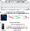
Figure 3. Identification of a KMT2A-ARHGEF12 fusion gene generated by an interstitial deletion in subband 11q23.3. (A) Array comparative genome hybridization detects a deletion which starts in the KMT2A gene and ends in the ARHGEF12 gene. (B) Fluorescence in situ hybridization confirms the deletion between the KMT2A and ARHGEF12 genes. The green and red probes hybridized on the normal chromosome 11. Only the red probes hybridized on the del(11) indicating the deleted area (green signals missing). (C) Examinations using molecular methodologies (genomic PCR and Sanger sequencing of the PCR amplified fragments) show that an intronic sequence of KMT2A fuses to an intronic sequence of ARHGEF12, generating a chimeric KMT2A-ARHGEF12 gene. Data and figure are from reference 305

References

    1. Rabbitts TH. Chromosomal translocations in human cancer. Nature. 1994;372(6502):143–149. doi: 10.1038/372143a0. - DOI - PubMed
    1. Rowley JD. The critical role of chromosome translocations in human leukemias. Annu Rev Genet. 1998;32:495–519. doi: 10.1146/annurev.genet.32.1.495. - DOI - PubMed
    1. Mitelman F, Johansson B, Mertens F. The impact of translocations and gene fusions on cancer causation. Nat Rev Cancer. 2007;7(4):233–245. doi: 10.1038/nrc2091. - DOI - PubMed
    1. Edwards PA. Fusion genes and chromosome translocations in the common epithelial cancers. J Pathol. 2010;220(2):244–254. doi: 10.1002/path.2632. - DOI - PubMed
    1. Zheng J. Oncogenic chromosomal translocations and human cancer (review) Oncol Rep. 2013;30(5):2011–2019. doi: 10.3892/or.2013.2677. - DOI - PubMed