

Multiple substrate recognition by yeast diadenosine and diphosphoinositol polyphosphate phosphohydrolase through phosphate clamping
- PMID: 33893105
- PMCID: PMC8064635
- DOI: 10.1126/sciadv.abf6744
Multiple substrate recognition by yeast diadenosine and diphosphoinositol polyphosphate phosphohydrolase through phosphate clamping
Abstract
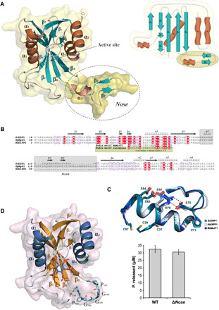
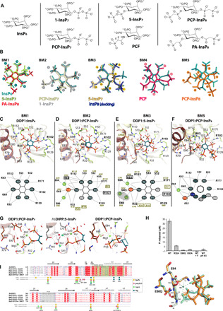
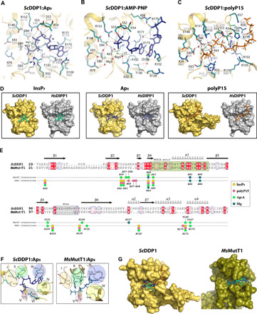
The yeast diadenosine and diphosphoinositol polyphosphate phosphohydrolase DDP1 is a Nudix enzyme with pyrophosphatase activity on diphosphoinositides, dinucleotides, and polyphosphates. These substrates bind to diverse protein targets and participate in signaling and metabolism, being essential for energy and phosphate homeostasis, ATPase pump regulation, or protein phosphorylation. An exhaustive structural study of DDP1 in complex with multiple ligands related to its three diverse substrate classes is reported. This allowed full characterization of the DDP1 active site depicting the molecular basis for endowing multisubstrate abilities to a Nudix enzyme, driven by phosphate anchoring following a defined path. This study, combined with multiple enzyme variants, reveals the different substrate binding modes, preferences, and selection. Our findings expand current knowledge on this important structural superfamily with implications extending beyond inositide research. This work represents a valuable tool for inhibitor/substrate design for ScDDP1 and orthologs as potential targets to address fungal infections and other health concerns.
Copyright © 2021 The Authors, some rights reserved; exclusive licensee American Association for the Advancement of Science. No claim to original U.S. Government Works. Distributed under a Creative Commons Attribution License 4.0 (CC BY).
Figures

Similar articles
-
Site-directed mutagenesis of diphosphoinositol polyphosphate phosphohydrolase, a dual specificity NUDT enzyme that attacks diadenosine polyphosphates and diphosphoinositol polyphosphates.J Biol Chem. 1999 Dec 10;274(50):35434-40. doi: 10.1074/jbc.274.50.35434. J Biol Chem. 1999. PMID: 10585413
-
Nudix hydrolases that degrade dinucleoside and diphosphoinositol polyphosphates also have 5-phosphoribosyl 1-pyrophosphate (PRPP) pyrophosphatase activity that generates the glycolytic activator ribose 1,5-bisphosphate.J Biol Chem. 2002 Dec 6;277(49):47313-7. doi: 10.1074/jbc.M209795200. Epub 2002 Oct 4. J Biol Chem. 2002. PMID: 12370170
-
Polyphosphates and Polyphosphatase Activity in the Yeast Saccharomyces cerevisiae during Overexpression of the DDP1 Gene.Biochemistry (Mosc). 2015 Oct;80(10):1312-7. doi: 10.1134/S0006297915100120. Biochemistry (Mosc). 2015. PMID: 26567575
-
Diphosphoinositol polyphosphates: the final frontier for inositide research?Biol Chem. 1999 Jul-Aug;380(7-8):945-51. doi: 10.1515/BC.1999.117. Biol Chem. 1999. PMID: 10494846 Review.
-
pH homeostasis in yeast; the phosphate perspective.Curr Genet. 2018 Feb;64(1):155-161. doi: 10.1007/s00294-017-0743-2. Epub 2017 Aug 30. Curr Genet. 2018. PMID: 28856407 Free PMC article. Review.
Cited by
-
Biochemical and structural characterization of an inositol pyrophosphate kinase from a giant virus.EMBO J. 2024 Feb;43(3):462-480. doi: 10.1038/s44318-023-00005-0. Epub 2024 Jan 12. EMBO J. 2024. PMID: 38216735 Free PMC article.
-
Activities and genetic interactions of fission yeast Aps1, a Nudix-type inositol pyrophosphatase and inorganic polyphosphatase.mBio. 2024 Aug 14;15(8):e0108424. doi: 10.1128/mbio.01084-24. Epub 2024 Jun 28. mBio. 2024. PMID: 38940614 Free PMC article.
-
Inositol Pyrophosphate-Controlled Kinetochore Architecture and Mitotic Entry in S. pombe.J Fungi (Basel). 2022 Sep 2;8(9):933. doi: 10.3390/jof8090933. J Fungi (Basel). 2022. PMID: 36135658 Free PMC article.
-
Duf89 abets lncRNA control of fission yeast phosphate homeostasis via its antagonism of precocious lncRNA transcription termination.RNA. 2023 Jun;29(6):808-825. doi: 10.1261/rna.079595.123. Epub 2023 Mar 7. RNA. 2023. PMID: 36882296 Free PMC article.
-
Cleavage-Polyadenylation Factor Cft1 and SPX Domain Proteins Are Agents of Inositol Pyrophosphate Toxicosis in Fission Yeast.mBio. 2022 Feb 22;13(1):e0347621. doi: 10.1128/mbio.03476-21. Epub 2022 Jan 11. mBio. 2022. PMID: 35012333 Free PMC article.
References
Publication types
Grants and funding
LinkOut - more resources
Full Text Sources
Other Literature Sources
Molecular Biology Databases
