. 2021 Jun 11;87(13):e0042321.
doi: 10.1128/AEM.00423-21. Epub 2021 Jun 11.

Manipulating Interactions between T4 Phage Long Tail Fibers and Escherichia coli Receptors

Affiliations

Manipulating Interactions between T4 Phage Long Tail Fibers and Escherichia coli Receptors

Akiyo Suga et al. Appl Environ Microbiol. .

Abstract

Bacteriophages are the most abundant and diverse biological entities on Earth. Phages exhibit strict host specificity that is largely conferred by adsorption. However, the mechanism underlying this phage host specificity remains poorly understood. In this study, we examined the interaction between outer membrane protein C (OmpC), one of the Escherichia coli receptors, and the long tail fibers of bacteriophage T4. T4 phage uses OmpC of the K-12 strain, but not of the O157 strain, for adsorption, even though OmpCs from the two E. coli strains share 94% homology. We identified amino acids P177 and F182 in loop 4 of the K-12 OmpC as essential for T4 phage adsorption in the copresence of loops 1 and 5. Analyses of phage mutants capable of adsorbing to OmpC mutants demonstrated that amino acids at positions 937 and 942 of the gp37 protein, which is present in the distal tip (DT) region of the T4 long tail fibers, play an important role in adsorption. Furthermore, we created a T4 phage mutant library with artificial modifications in the DT region and isolated and characterized multiple phage mutants capable of adsorbing to OmpC of the O157 strain or lipopolysaccharide of the K-12 strain. These results shed light on the mechanism underlying the phage host specificity mediated by gp37 and OmpC and may be useful in the development of phage therapy via artificial modifications of the DT region of T4 phage. IMPORTANCE Understanding the host specificity of phages will lead to the development of phage therapy. The interaction between outer membrane protein C (OmpC), one of the Escherichia coli receptors, and the gp37 protein present in the distal tip (DT) region of the long tail fibers of T4 bacteriophages largely determines their host specificity. Here, we elucidated the amino acid residues important for the interaction between gp37 and OmpC. This result suggests that the shapes of both proteins at the binding interface play important roles in their interactions, which are likely mediated by multiple residues of both binding partners. Additionally, we successfully isolated multiple phage mutants capable of adsorbing to a variety of E. coli receptors using a mutant T4 phage library with artificial modifications in the DT region, providing a foundation for the alteration of the host specificity.

Keywords: Escherichia coli; bacteriophage therapy; bacteriophages.


Figures

FIG 1
FIG 1
Growth and adsorption of T4 phage on cells expressing OmpCK12 or OmpCO157. (A) The suspension containing the number of T4 phages indicated on the left was applied as spots onto an LB plate containing 0.2% l-Ara seeded with TY0807, O157:H7, TY0807 ΔompC, and TY0807 ΔompC harboring pBAD33-OmpCK12 or pBAD33-OmpCO157, and the plates were incubated overnight at 37°C. “–” indicates no plasmid. TY0807 was used as a wild-type Escherichia coli K-12 strain. (B) Adsorption analyses were performed as described in Materials and Methods. Symbols: ○, TY0807; □, O157:H7; ●, TY0807 ΔompC; ▴, TY0807 ΔompC harboring pBAD33-OmpCK12; ◆, TY0807 ΔompC harboring pBAD33-OmpCO157. The number of unadsorbed phages was determined by plating with TY0807 as an indicator.
FIG 2
FIG 2
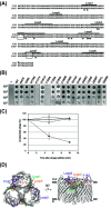
Growth and adsorption of T4 phage on cells expressing OmpC mutants. (A) Alignment of amino acid sequence in OmpC from Escherichia coli strains K-12 and O157. The lines or the asterisks indicate amino acids forming extracellular loop structures in OmpCK12 or different amino acids between OmpCK12 and OmpCO157. The black boxes represent a 4-amino-acid insertion specific to OmpCK12 or OmpCO157. (B) The suspension containing the number of T4 phages indicated on the left was applied as spots onto an LB plate containing 0.2% l-Ara seeded with TY0807 ΔompC harboring each plasmid expressing the OmpC mutants indicated on the top, and the plates were incubated overnight at 37°C. “–” indicates no plasmid. TY0807 was used as a wild-type E. coli K-12 strain. (C) Adsorption analyses were performed as described in Materials and Methods. Symbols: ○, TY0807 ΔompC harboring pBAD33; ●, TY0807 ΔompC harboring pBAD33-OmpCK12; ◇, TY0807 ΔompC harboring pBAD33-OmpCK12(P177V); ◆, TY0807 ΔompC harboring pBAD33-OmpCK12(F182A). The number of unadsorbed phages was determined by plating with TY0807 as an indicator. (D) OmpC viewed from the top (left) and the side (right) is shown (PDB ID 2J1N). Proline at position 177 (red), phenylalanine at position 182 (blue), and loops 1 (green), 4 (orange), and 5 (purple) are highlighted. Gray bars indicate a predicted membrane boundary.
FIG 3
FIG 3
Growth and adsorption of T4 phage on cells expressing chimeric OmpCO157 with OmpCK12 loops. (A) Schematic diagram of chimeric OmpC mutants. The lines above OmpCK12 indicate regions of extracellular loop structures in OmpC. (B) The suspension containing the number of T4 phages indicated on the left was applied as spots onto an LB plate containing 0.2% l-Ara seeded with TY0807, TY0807 ΔompC, or TY0807 ΔompC harboring pBAD33-ompCK12, pBAD33-OmpCO157, pBAD33-OmpCO157ΔGVIN, pBAD33-OmpCO157ΔGVIN+K12Loop4, pBAD33-OmpCO157ΔGVIN+K12Loop4,5, or pBAD33-OmpCO157ΔGVIN+K12Loop1,4,5, and the plates were incubated overnight at 37°C. “–” indicates no plasmid. TY0807 was used as a wild-type Escherichia coli K-12 strain. (C) Adsorption analyses were performed as described in Materials and Methods. Symbols: ○, TY0807; ●, TY0807 ΔompC; △, TY0807 ΔompC harboring pBAD33-OmpCK12; ▴, TY0807 ΔompC harboring pBAD33-OmpCO157; □, TY0807 ΔompC harboring pBAD33-OmpCO157ΔGVIN; ■, TY0807 ΔompC harboring pBAD33-OmpCO157ΔGVIN+K12Loop4; ◇, TY0807 ΔompC harboring pBAD33-OmpCO157ΔGVIN+K12Loop4,5; ◆, TY0807 ΔompC harboring pBAD33-OmpCO157ΔGVIN+K12Loop1,4,5. The number of unadsorbed phages was determined by plating with TY0807 as an indicator.
FIG 4
FIG 4
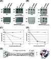
Characterization of T4 phage mutants capable of growing on cells expressing OmpCK12(P177V) or OmpCK12(F182A). (A) The suspensions containing the number of wild-type (WT) or mutant (M1) T4 phages indicated on the left were applied as spots onto an LB plate containing 0.2% l-Ara seeded with TY0807, TY0807 ΔompC, and TY0807 ΔompC harboring pBAD33-OmpCK12 or pBAD33-OmpCK12(P177V), as indicated on the bottom, and the plates were incubated overnight at 37°C. (B) The suspensions containing the number of WT or mutant (M2) T4 phages indicated on the left were applied as spots onto an LB plate containing 0.2% l-Ara seeded with TY0807, TY0807 ΔompC, and TY0807 ΔompC harboring pBAD33-OmpCK12 or pBAD33-OmpCK12(F182A), as indicated on the bottom, and the plates were incubated overnight at 37°C. (C) Adsorption analyses of T4 phage mutant (N937S) were performed as described in Materials and Methods. Symbols: ●, TY0807 ΔompC harboring pBAD33; ■, TY0807 ΔompC harboring pBAD33-OmpCK12; ▴, TY0807 ΔompC harboring pBAD33-OmpCK12(P177V). The number of unadsorbed phages was determined by plating with TY0807 as an indicator. (D) Adsorption analyses of T4 phage mutant (G942R) were performed as described in Materials and Methods. Symbols: ●, TY0807 ΔompC harboring pBAD33; ■, TY0807 ΔompC harboring pBAD33-OmpCK12; ▴, TY0807 ΔompC harboring pBAD33-OmpCK12(F182A). The number of unadsorbed phages was determined by plating with TY0807 as an indicator. (E) The DT region of T4 phage long tail fiber (gp37) viewed from the side (left) and the top (right) is shown (PDB ID 2XGF). Note that the top view is enlarged. N937 (red) and G942 (blue) are highlighted.
FIG 5
FIG 5
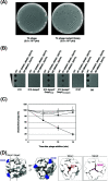
Isolation and characterization of T4 phage mutants capable of growing on cells expressing OmpCO157. (A) The phage library (2 × 1010 PFU) prepared with regular PCR (left plate) or error-prone PCR (right plate) was plated onto an LB plate containing 0.2% l-Ara seeded with TY0807 ΔompC harboring pBAD33-OmpCO157 and incubated overnight at 37°C. (B) The suspensions containing the number of wild-type (WT) or mutant (M1 and M2) T4 phages shown (105, 103, or 101 PFU for wild type and M1 and 103, 102, or 101 PFU for M2) were applied as spots onto an LB plate containing 0.2% l-Ara seeded with TY0807, TY0807 ΔompC, TY0807 ΔompC harboring pBAD33-OmpCO157 or pBAD33-OmpCO157ΔGVIN, O157:H7, or BB, as indicated on the bottom, and the plates were incubated overnight at 37°C. (C) Adsorption analyses of T4 phage mutant (G940V, M946V, and S947P) were performed as described in Materials and Methods. Symbols: ●, TY0807; ○, TY0807 ΔompC; ■, TY0807 ΔompC harboring pBAD33-OmpCO157; □, O157:H7. The number of unadsorbed phages was determined by plating with TY0807 ΔompC harboring pBAD33-OmpCO157 as an indicator. (D) Top views of the DT region of gp37 showing the surface (left) and sliced interiors (right) (PDB ID 2XGF). G940 (blue), M946 (pink), and S947 (red) are highlighted.
FIG 6
FIG 6
Isolation and characterization of the T4 phage mutants capable of growing on ΔompC cells. (A) The phage library (2 × 1010 PFU) prepared with regular PCR (left plate) or error-prone PCR (right plate) was plated onto an LB plate seeded with TY0807 ΔompC cells and incubated overnight at 37°C. (B) The suspensions containing the different numbers (101, 102, 103, and 104) of wild-type (WT) or mutant (M1 and M2) T4 phages indicated on the top were applied as spots onto an LB plate seeded with BB, TY0807, TY0807 ΔompC, TY0726, or TY0728, and the plates were incubated overnight at 37°C. TY0807 or BB was used as a wild-type Escherichia coli K-12 strain or B strain, respectively. (C) Top views of the DT region of gp37 showing the surface (PDB ID 2XGF). S952 (blue) and Y953 (red) are highlighted.
FIG 7
FIG 7
DT-OmpC docking model. The DT region of gp37 (blue [modified from PDB ID 2XGF]) was docked with OmpC (white [PDB ID 2J1N]) using the ClusPro server. Gray bars indicate the predicted membrane boundary.

Similar articles

Cited by

References

    1. Suttle CA. 2007. Marine viruses—major players in the global ecosystem. Nat Rev Microbiol 5:801–812. 10.1038/nrmicro1750. - DOI - PubMed
    1. Miller EM, Goebel WF. 1949. Studies on bacteriophage. I. The relationship between the somatic antigens of Shigella sonnei and their susceptibility to bacterial viruses. J Exp Med 90:255–265. 10.1084/jem.90.3.255. - DOI - PMC - PubMed
    1. Jesaitis MA, Goebel WF. 1953. The interaction between T4 phage and the specific lipocarbohydrate of phase II Sh. sonnei. Cold Spring Harbor Symp Quant Biol 18:205–208. 10.1101/SQB.1953.018.01.031. - DOI - PubMed
    1. Goldberg E, Grinius L, Letellier L. 1994. Recognition, attachment, and infection, p 347–356. In Karam JD, Drake JW, Kreuzer KN, Mosig G, Hall DH, Eisering FA, Black LW, Spicer EK, Kutter E, Carlson K, Miller ES (ed), Molecular biology of bacteriophage T4, vol 1. ASM Press, Washington, DC.
    1. Simon LD, Anderson TF. 1967. The infection of Escherichia coli by T2 and T4 bacteriophages as seen in the electron microscope. I. Attachment and penetration. Virology 32:279–297. 10.1016/0042-6822(67)90277-2. - DOI - PubMed

Publication types

MeSH terms

LinkOut - more resources