. 2021 Jun 11;87(13):e0044221.
doi: 10.1128/AEM.00442-21. Epub 2021 Jun 11.

Probiotic Bacillus Affects Enterococcus faecalis Antibiotic Resistance Transfer by Interfering with Pheromone Signaling Cascades

Affiliations

Probiotic Bacillus Affects Enterococcus faecalis Antibiotic Resistance Transfer by Interfering with Pheromone Signaling Cascades

Yu-Chieh Lin et al. Appl Environ Microbiol. .

Abstract

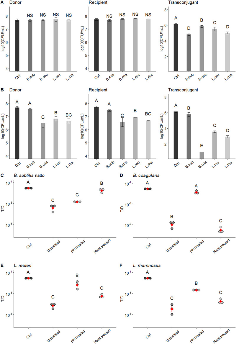
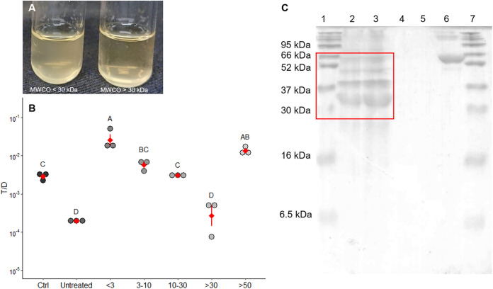
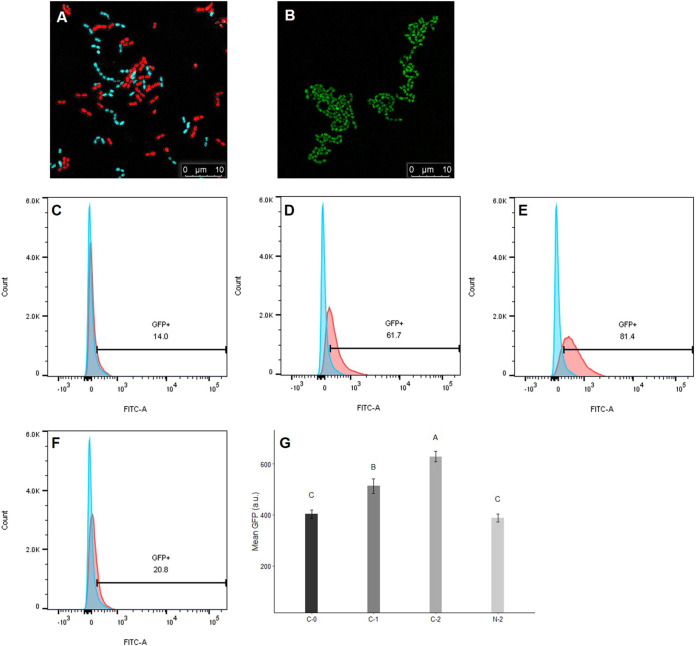
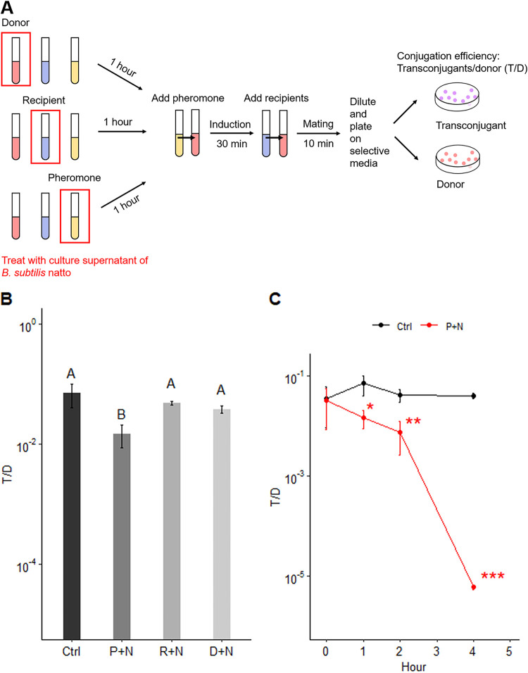
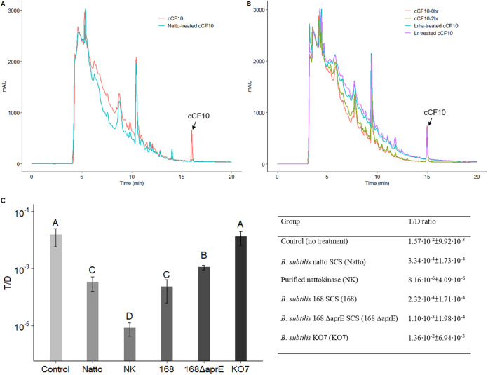
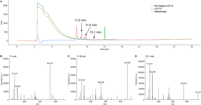
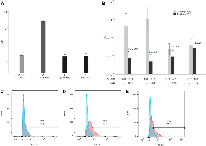
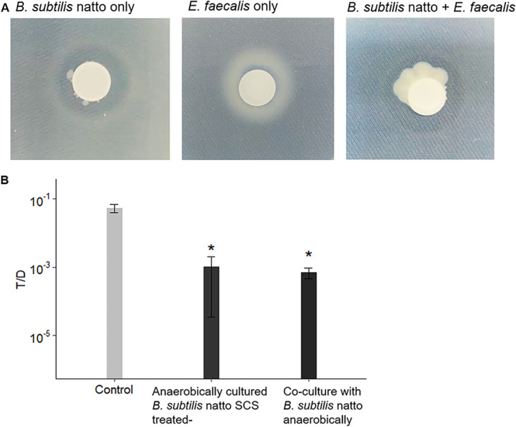
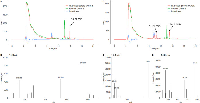
Enterococcus faecalis, a member of the commensal flora in the human gastrointestinal tract, has become a threatening nosocomial pathogen because it has developed resistance to many known antibiotics. More concerningly, resistance gene-carrying E. faecalis cells may transfer antibiotic resistance to resistance-free E. faecalis cells through their unique quorum sensing-mediated plasmid transfer system. Therefore, we investigated the role of probiotic bacteria in the transfer frequency of the antibiotic resistance plasmid pCF10 in E. faecalis populations to mitigate the spread of antibiotic resistance. Bacillus subtilis subsp. natto is a probiotic strain isolated from Japanese fermented soybean foods, and its culture fluid potently inhibited pCF10 transfer by suppressing peptide pheromone activity from chromosomally encoded CF10 (cCF10) without inhibiting E. faecalis growth. The inhibitory effect was attributed to at least one 30- to 50-kDa extracellular protease present in B. subtilis subsp. natto. Nattokinase of B. subtilis subsp. natto was involved in the inhibition of pCF10 transfer and cleaved cCF10 (LVTLVFV) into LVTL plus VFV fragments. Moreover, the cleavage product LVTL (L peptide) interfered with the conjugative transfer of pCF10. In addition to cCF10, faecalis-cAM373 and gordonii-cAM373, which are mating inducers of vancomycin-resistant E. faecalis, were also cleaved by nattokinase, indicating that B. subtilis subsp. natto can likely interfere with vancomycin resistance transfer in E. faecalis. Our work shows the feasibility of applying fermentation products of B. subtilis subsp. natto and L peptide to mitigate E. faecalis antibiotic resistance transfer. IMPORTANCE Enterococcus faecalis is considered a leading cause of hospital-acquired infections. Treatment of these infections has become a major challenge for clinicians because some E. faecalis strains are resistant to multiple clinically used antibiotics. Moreover, antibiotic resistance genes can undergo efficient intra- and interspecies transfer via E. faecalis peptide pheromone-mediated plasmid transfer systems. Therefore, this study provided the first experimental demonstration that probiotics are a feasible approach for interfering with conjugative plasmid transfer between E. faecalis strains to stop the transfer of antibiotic resistance. We found that the extracellular protease(s) of Bacillus subtilis subsp. natto cleaved peptide pheromones without affecting the growth of E. faecalis, thereby reducing the frequency of conjugative plasmid transfer. In addition, a specific cleaved pheromone fragment interfered with conjugative plasmid transfer. These findings provide a potential probiotic-based method for interfering with the transfer of antibiotic resistance between E. faecalis strains.

Keywords: Bacillus subtilis subsp. natto; Enterococcus faecalis; antibiotic resistance; nattokinase; pheromone-inducible conjugative plasmid transfer; probiotics.


Figures

FIG 1
FIG 1
Effect of pH-treated or heat-treated probiotic spent culture supernatants on growth and pCF10 transfer of E. faecalis. Spent culture supernatants (SCS) of aerobically cultured B. subtilis subsp. natto, B. coagulans, L. reuteri, and L. rhamnosus were first treated with pH adjustment (pH adjusted to 6.5 with NaOH) or heat (80°C water bath for 30 min) and were then added to the E. faecalis mating culture. After a 4-h mating assay, the concentrations of donors, recipients, and transconjugants were determined by plate counting. (A) Effect of pH-adjusted probiotic SCSs. (B) Effect of heat-treated probiotic SCSs. In panels A and B, the concentrations of donors, recipients, and transconjugants are presented as log10(CFU/ml) values. The data are presented as the means ± standard deviations (SDs) (n = 3). (C to F) Effects of untreated, pH-treated, or heat-treated probiotic SCSs on the number of transconjugants per donor (T/D ratios). Each dot in the figure represents a replication. The red diamonds with red lines indicate the means ± SDs (n = 3). Multiple comparisons were evaluated using Duncan’s multiple-range test. The different uppercase letters above the bars or dots indicate the significance of differences between groups (P < 0.05). Ctrl, negative control (the group treated with fresh M9B medium; no SCS treatment); B. sub, B. subtilis subsp. natto; B. coa, B. coagulans; L. reu, L. reuteri; L. rha, L. rhamnosus; NS, no significant difference.
FIG 2
FIG 2
At least one 30- to 50-kDa protein in B. subtilis subsp. natto supernatant inhibits pCF10 transfer. (A) Mating culture of E. faecalis treated with different MW fractions of B. subtilis subsp. natto supernatant. In this picture, the turbidity of the culture on the left, which was treated with the fraction with a MW less than 30 kDa appears higher than that of the culture on the right, which was treated with the fraction with a MW greater than 30 kDa. (B) Effect of different MW fractions of B. subtilis subsp. natto supernatant on the conjugative transfer of pCF10. The y axis indicates the T/D ratio, and the x axis indicates different MW (kDa) fractions of B. subtilis subsp. natto supernatant. Ctrl, negative control (the group treated with fresh M9B medium; no SCS treatment); untreated, the group treated with unfractionated B. subtilis subsp. natto supernatant. Each dot in the figure represents a replication. The red diamonds with red lines indicate the means ± SDs (n = 3). Multiple comparisons were evaluated using Duncan’s multiple-range test. The different uppercase letters above the dots indicate the significance of differences between groups (P < 0.05). (C) SDS-PAGE of B. subtilis subsp. natto supernatant. Lanes 1 and 7, protein marker; lane 2, B. subtilis subsp. natto supernatant (5 μl); lane 3, B. subtilis subsp. natto supernatant (10 μl); lane 4, M9B medium (5 μl); lane 5, M9B medium (10 μl); lane 6, BSA.
FIG 3
FIG 3
Influence of B. subtilis subsp. natto supernatant on the cCF10-inducible GFP reporter harbored on pCF10. (A) Under confocal microscopy, E. faecalis OG1Sp/pCF10-iGFP-p23-tdTomato (donor) appears red, and E. faecalis OG1RF::p23cfp (recipient) appears cyan. In the middle of the image, donor and recipient cells were close together, indicating that mating was likely happening. (B) When donor cells were induced with cCF10, they expressed the GFP reporter and exhibited green aggregates. (C to F) Donor cells were induced with 2.5 ng/ml cCF10 and harvested at 0, 60, and 120 min. Next, the expression of the GFP reporter was quantified using flow cytometry. Blue peak, wild-type E. faecalis OG1RF that would not express the GFP reporter, used as the negative control. Red peaks, donor cells induced with 0 ng/ml cCF10 for 0 min (C), 2.5 ng/ml cCF10 for 60 min (D), 2.5 ng/ml cCF10 for 120 min (E), or 2.5 ng/ml cCF10 plus B. subtilis subsp. natto SCS for 120 min (F). The numbers shown in the middle of panels C to F are the percentages of GFP-positive cells after gating. (G) Mean GFP intensity of GFP-positive donor cells after gating. Donor cells were induced with 0 ng/ml cCF10 for 0 min (C-0), 2.5 ng/ml cCF10 for 60 min (C-1), 2.5 ng/ml cCF10 for 120 min (C-2), or 2.5 ng/ml cCF10 plus B. subtilis subsp. natto SCS for 120 min (N-2). The data are presented as the means ± SDs (n = 3). Values with different uppercase letters were significantly different according to Duncan’s multiple-range tests (P < 0.05).
FIG 4
FIG 4
B. subtilis subsp. natto supernatant affects the activity of cCF10. (A) Modified E. faecalis mating experiment. The SCS of B. subtilis subsp. natto was first treated separately with donor cells, recipient cells, or pheromone cCF10 at 37°C for 1 h. Next, donor cells were incubated with cCF10 at 37°C for 30 min and then mixed with recipient cells for one round of conjugation (10 min). Through this method, we were able to clarify the factor that is actually affected by the SCS of B. subtilis subsp. natto. (B) T/D ratios after separate treatment of pheromone cCF10, recipient cells, or donor cells with B. subtilis subsp. natto SCS. The y axis indicates the T/D ratio. Ctrl (control), not treated with SCS, P+N: pheromone cCF10 treated with B. subtilis subsp. natto SCS; R+N, recipient cells treated with B. subtilis subsp. natto SCS; D+N, donor cells treated with B. subtilis subsp. natto SCS. The data are presented as the means ± SDs (n = 3). Values with different uppercase letters were significantly different according to Duncan’s multiple-range tests (P < 0.05). (C) The effect of B. subtilis subsp. natto SCS on cCF10 increased over time. Donor cells were induced with cCF10 that was first treated with B. subtilis subsp. natto SCS for 0, 1, 2, and 4 h. Then, recipient cells were added and allowed one round of conjugation. The y axis indicates the T/D ratio. Ctrl (control), not treated with SCS; P+N, pheromone cCF10 treated with B. subtilis subsp. natto SCS. The data are presented as the means ± SDs (n = 3). *, P < 0.05; **, P < 0.01; ***, P < 0.001 compared with each negative control group (Student’s t test).
FIG 5
FIG 5
An extracellular protease of B. subtilis subsp. natto cleaves cCF10 and inhibits pCF10 transfer. (A and B) HPLC chromatograms of probiotic supernatant-treated peptide pheromone cCF10. The synthetic peptide pheromone cCF10 (100 ng/ml) dissolved in M9B medium (150 μl total) was treated with 50 μl of probiotic SCSs or M9B medium at 37°C for 30 min. Next, the assay mixtures were analyzed using HPLC. The peak of cCF10 is at 15.2 min. (A) cCF10 was degraded when treated with B. subtilis subsp. natto SCS (Natto). (B) cCF10 was not degraded in M9B medium over time (0 to 2 h) or when treated with the SCS of L. rhamnosus (Lrha) or L. reuteri (Lr). (C) Conjugation assays with exogenously added B. subtilis 168 ΔaprE SCS, B. subtilis KO7 SCS, and purified nattokinase (final conc., 1 mg/ml). Donor and recipient cells were mixed 1:1 in M9B medium with and without treatment described above and allowed to grow for 4 h. The entire culture was then plated on selective medium for transconjugants and donors. The y axis indicates the T/D ratio. The data are presented as the means ± SDs (n = 3). Values with different uppercase letters were significantly different according to Duncan’s multiple-range tests (P < 0.05).
FIG 6
FIG 6
HPLC chromatogram and mass spectra of nattokinase-treated cCF10. The synthetic peptide pheromone cCF10 (100 ng/ml) dissolved in 50 mM phosphate buffer containing 150 mM NaCl (pH 7.0) (150 μl total) was treated with 50 μl of 1 mg/ml B. subtilis nattokinase solution at 37°C for 30 min. Next, the assay mixtures were analyzed using HPLC and MS. (A) The peak of cCF10 is at 15.2 min. After cCF10 was treated with nattokinase (NK), the reaction product was found to produce three new peaks, as shown in the HPLC chromatogram (RTs, 11.0, 11.6, and 13.1 min). The mass spectra of these three new peaks are shown in panels B to D.
FIG 7
FIG 7
Influence of L peptide on the conjugative transfer of pCF10. Donor cells were incubated with L peptide (L) (cCF10 fragment, sequence LVTL) alone or together with cCF10 (C) at 37°C for 30 min. Then, recipient cells were added and incubated for 10 min to allow one round of conjugation before the entire culture was plated on selective medium for transconjugants and donors. The conjugation frequency is represented by the number of transconjugants per donor (T/D ratio). (A) L was added at physiological (3.16 nM) or higher concentrations (15.8 nM) to induce donor cells. The groups induced with no peptide (L or C) or 3.16 nM C served as the negative-control and positive-control groups, respectively. The data are presented as the means ± SDs (n = 3). (B) Different volumes of 1 mg/ml L stock solution (dissolved in dimethyl sulfoxide [DMSO]) and a fixed volume of 1 mg/ml C stock solution (dissolved in DMSO; final conc., 3.16 nM) were added to achieve L/C ratios of 0.25:1, 0.5:1, 1:1, and 2:1 for induction of donor cells (treatment groups). The groups induced with 3.16 nM C served as the negative control. In addition, to eliminate the effect of DMSO, a volume of DMSO equal to that of the L stock solution was added to the negative control group. The data are presented as the means ± SDs (n = 3). *, P < 0.05; **, P < 0.01 compared with each negative control group (Student’s t test). (C to E) The expression of the cCF10-inducible GFP reporter harbored on pCF10 was quantified using flow cytometry. Blue peak, wild-type E. faecalis OG1RF that would not express the GFP reporter was used as the negative control. Red peaks, donor cells induced with no peptides for 0 min (C), 0.7 μl DMSO plus 3.16 nM C for 60 min (D), or 1.58 nM L plus 3.16 nM C (L/C = 1:2) for 60 min (E). The numbers shown in the middles of panels C to E are the percentages of GFP-positive cells after gating.
FIG 8
FIG 8
B. subtilis subsp. natto grows and secretes proteases under anaerobic conditions. (A) B. subtilis subsp. natto was cultured alone or together with E. faecalis donors and recipients on SGN (Schaeffer’s sporulation medium supplemented with glucose and nitrate) agar supplemented with skim milk. The amount of secreted protease was estimated from the clear zone that formed around the filter membrane. (B) Four-hour in vitro conjugation assays with exogenously added anaerobically cultured B. subtilis subsp. natto supernatant (SGN broth, 48 h) or B. subtilis subsp. natto (coculture). For bacterial coculture experiments, the cultures of B. subtilis subsp. natto were grown anaerobically in SGN broth to the early exponential phase and diluted according to the OD600. Next, the cultures were added to E. faecalis mating cultures and allowed to grow for 4 h. The entire culture was then plated on selective medium for E. faecalis transconjugants and donors. Fresh SGN broth was added to mating cultures to serve as the negative control group. The y axis indicates the T/D ratio. The data are presented as the means ± SDs (n = 3). *, P < 0.05 compared with negative control (Duncan’s multiple-range tests).
FIG 9
FIG 9
HPLC chromatogram and mass spectra of nattokinase-treated faecalis-cAM373 and gordonii-cAM373. The synthetic peptide pheromone (faecalis-cAM373 or gordonii-cAM373) (100 ng/ml) dissolved in 50 mM phosphate buffer containing 150 mM NaCl (pH 7.0) (150 μl total) was treated with 50 μl of 1 mg/ml B. subtilis nattokinase solution at 37°C for 30 min. Next, the assay mixtures were analyzed using HPLC and MS. (A) The peak of faecalis-cAM373 (sequence AIFILAS) occurred at 13.5 min. After faecalis-cAM373 was treated with nattokinase (NK), the reaction product was found to produce a new peak, as shown in the HPLC chromatogram (RT, 14.9 min). The mass spectrum of this new peak is shown in panel B. (C) The peak of gordonii-cAM373 (sequence SVFILAA) occurred at 13.6 min. After gordonii-cAM373 was treated with nattokinase (NK), the reaction product was found to produce two new peaks, as shown in the HPLC chromatogram (RTs, 10.1 and 14.2 min). The mass spectra of these two new peaks are shown in panels D and E.

Similar articles

Cited by

References

    1. Paulsen IT, Banerjei L, Myers GS, Nelson KE, Seshadri R, Read TD, Fouts DE, Eisen JA, Gill SR, Heidelberg JF, Tettelin H, Dodson RJ, Umayam L, Brinkac L, Beanan M, Daugherty S, DeBoy RT, Durkin S, Kolonay J, Madupu R, Nelson W, Vamathevan J, Tran B, Upton J, Hansen T, Shetty J, Khouri H, Utterback T, Radune D, Ketchum KA, Dougherty BA, Fraser CM. 2003. Role of mobile DNA in the evolution of vancomycin-resistant Enterococcus faecalis. Science 299:2071–2074. 10.1126/science.1080613. - DOI - PubMed
    1. Aakra A, Vebø H, Snipen L, Hirt H, Aastveit A, Kapur V, Dunny G, Murray BE, Murray B, Nes IF. 2005. Transcriptional response of Enterococcus faecalis V583 to erythromycin. Antimicrob Agents Chemother 49:2246–2259. 10.1128/AAC.49.6.2246-2259.2005. - DOI - PMC - PubMed
    1. Hanchi H, Mottawea W, Sebei K, Hammami R. 2018. The genus Enterococcus: between probiotic potential and safety concerns-an update. Front Microbiol 9:1791. 10.3389/fmicb.2018.01791. - DOI - PMC - PubMed
    1. Lindenstrauss AG, Ehrmann MA, Behr J, Landstorfer R, Haller D, Sartor RB, Vogel RF. 2014. Transcriptome analysis of Enterococcus faecalis toward its adaption to surviving in the mouse intestinal tract. Arch Microbiol 196:423–433. 10.1007/s00203-014-0982-2. - DOI - PubMed
    1. Jett BD, Huycke MM, Gilmore MS. 1994. Virulence of enterococci. Clin Microbiol Rev 7:462–478. 10.1128/cmr.7.4.462. - DOI - PMC - PubMed

Publication types

LinkOut - more resources