. 2021 Apr 27;118(17):e2026102118.
doi: 10.1073/pnas.2026102118.

Probing the binding specificities of human Siglecs by cell-based glycan arrays

Affiliations

Probing the binding specificities of human Siglecs by cell-based glycan arrays

Christian Büll et al. Proc Natl Acad Sci U S A. .

Abstract

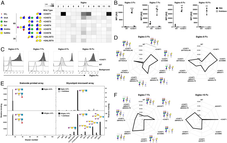
Siglecs are a family of sialic acid-binding receptors expressed by cells of the immune system and a few other cell types capable of modulating immune cell functions upon recognition of sialoglycan ligands. While human Siglecs primarily bind to sialic acid residues on diverse types of glycoproteins and glycolipids that constitute the sialome, their fine binding specificities for elaborated complex glycan structures and the contribution of the glycoconjugate and protein context for recognition of sialoglycans at the cell surface are not fully elucidated. Here, we generated a library of isogenic human HEK293 cells with combinatorial loss/gain of individual sialyltransferase genes and the introduction of sulfotransferases for display of the human sialome and to dissect Siglec interactions in the natural context of glycoconjugates at the cell surface. We found that Siglec-4/7/15 all have distinct binding preferences for sialylated GalNAc-type O-glycans but exhibit selectivity for patterns of O-glycans as presented on distinct protein sequences. We discovered that the sulfotransferase CHST1 drives sialoglycan binding of Siglec-3/8/7/15 and that sulfation can impact the preferences for binding to O-glycan patterns. In particular, the branched Neu5Acα2-3(6-O-sulfo)Galβ1-4GlcNAc (6'-Su-SLacNAc) epitope was discovered as the binding epitope for Siglec-3 (CD33) implicated in late-onset Alzheimer's disease. The cell-based display of the human sialome provides a versatile discovery platform that enables dissection of the genetic and biosynthetic basis for the Siglec glycan interactome and other sialic acid-binding proteins.

Keywords: CD33; Siglecs; cell-based glycan array; sialome; sialyltransferase.


Conflict of interest statement

Competing interest statement: University of Copenhagen has filed a patent application on the cell-based display platform. GlycoDisplay ApS, Copenhagen, Denmark, has obtained a license to the field of the patent application. Y.N. and H.C. are cofounders of GlycoDisplay ApS and hold ownerships in the company.

Figures

Fig. 1.
Fig. 1.
Generation of the sialoglycan sublibraries. (A) Scheme showing the sublibrary approach for display of α2-3 and α2-6 sialic acid capping of galactose and display of sialylated O-glycans by combinatorial KO (Δ) of endogenous genes (capitalized and italicized) and individual KI (+) of sialyltransferase complementary DNA (capitalized and nonitalicized). Starting from HEKWT cells, ST6GAL1/2 were knocked out to delete α2-6-sialylation of galactose. Next, ST3GAL1/2/5 and ST3GAL3/4/6 were knocked out in sets or combined. ST3GAL1-6 KO was performed, thus deleting α2-3-sialylation capacity. In these empty cells, HEKΔSia, the deleted sialyltransferase genes were knocked in individually. HEKWT cells predictably express a mixture of mSTa (Neu5Acα2-3Galβ1-3GalNAcα1-O-Ser/Thr), dST (Neu5Acα2-3Galβ1-3[Neu5Acα2-6]GalNAcα1-O-Ser/Thr), and sialylated core2 structures. COSMC (or C1GALT1) was knocked out for display of the Tn (GalNAcα-O-Thr/Ser) antigen followed by KI of either ST6GALNAC1 or B3GNT6 yielding STn (Neu5Acα2-6GalNAcα-O-Thr/Ser) or core3 structures (GlcNAcβ1-3GalNAcα1-O-Ser/Thr), respectively. Alternatively, GCNT1 was knocked out resulting in display of a mixture of mSTa/dST and combined KO of ST6GALNAC2/3/4-produced mSTa. Further KO of ST3GAL1/2 produced the T antigen (Galβ1-3GalNAcα1-O-Ser/Thr), and individual KI of ST6GALNAC2/3/4 yielded cells with dST expression. Predicted sialoglycan structures are depicted according to the Symbol Nomenclature for Glycans (92). (B) Radar charts show binding of Pan-Lectenz (Sia), α2-3-Lectenz (α2-3Sia), or SNA-I (α2-6Sia) to the sialic acid capping sublibrary. Lectin binding was quantified by flow cytometry, and data are shown as mean fluorescence intensity (MFI) values from three independent experiments. (C) Scheme illustrating metabolic labeling of cells with Ac5SiaNPoc that is incorporated into cell surface glycans and conjugated to fluorescent azide-biotin using click chemistry. Bar diagram shows metabolic labeling of the capping sublibrary with 0 to 100 µM Ac5SiaNPoc as average MFI ± SEM of three independent experiments. (D) Radar charts show binding of Pan-Lectenz (Sia), VVA (Tn), and PNA (T) to the O-glycan sublibrary as representative MFI values of three independent experiments.
Fig. 2.
Fig. 2.
Siglec-4 fine binding dissection with cell-based glycan and mucin reporter display. (A) Radar charts show Siglec-4 Fc binding to sialic acid capping (Left) and O-glycan (Right) sublibrary as representative MFI values of three independent experiments. Predicted glycan epitopes and essential glycosyltransferase genes from analysis with GlycoRadar are illustrated. (B) Illustration of the mucin reporter with 6xHis, FLAG tag, and GFP, 200 amino acid–interchangeable mucin TR domain for expression in isogenic cells as membrane-bound or secreted form. Bar diagram shows Siglec-4 binding to HEKWT and HEKdST(KO GCNT1/ST6GALNAC2/3/4, KI ST6GALNAC4) cells transiently transfected with 41 different mucin and mucin-like protein reporters as MFI values from three independent experiments. (C) MUC1, GP1bα, and MUC2 dot blot overlaid with Siglec-4 Fc (above). Equal blotting was confirmed by overlay with anti-6xHis (below). Serial dilutions of the mucin reporters were blotted, and human Fc (hFc) was blotted as positive control. (D) Siglec-4 binding to O-glycoengineered cells expressing membrane MUC1 reporter is shown as radar chart.
Fig. 3.
Fig. 3.
Siglec-7 fine binding dissection with cell-based glycan and mucin reporter display. (A) Radar charts showing Siglec-7 Fc binding to sialic acid capping (Left) and O-glycan (Right) sublibrary as representative MFI values of three independent experiments. Predicted glycan epitopes and essential glycosyltransferase genes from analysis with GlycoRadar are illustrated. (B) Bar diagram shows Siglec-7 binding to HEKWT and HEKSTn (KO COSMC KI ST6GALNAC1) cells transiently transfected with 41 different mucin reporters as MFI values from three independent experiments. (C) MUC1, GP1bα, and MUC2 dot blot overlaid with Siglec-7 Fc (above) and anti-6xHis (below). Serial dilutions of the mucin reporters were blotted, and human Fc was blotted as positive control. (D) Siglec-7 binding to O-glycoengineered cells expressing membrane GP1bα reporter. MFI from three independent experiments is shown in the radar chart.
Fig. 4.
Fig. 4.
Siglec-15 fine binding dissection with cell-based glycan and mucin reporter display. (A) Radar charts showing Siglec-15 Fc binding to sialic acid capping (Left) and O-glycan (Right) sublibrary as representative MFI values of three independent experiments. Predicted glycan epitopes and essential glycosyltransferase genes from analysis with GlycoRadar are illustrated. (B) Bar diagram shows Siglec-15 binding to HEKWT and HEKSTn (KO COSMC KI ST6GALNAC1) cells transiently transfected with 41 different mucin reporters as MFI values from three independent experiments. (C) Radar chart presentation of Siglec-15 binding to membrane MUC13 reporter expressed in O-glycan sublibrary. (D) Secreted mucin reporters (MUC1, MUC2, MUC5AC, MUC7, MUC13, and MUC22) produced in engineered HEK cells with WT, Tn, STn, dST, and mSTa glycosylation were immobilized and overlaid with Siglec-15 or anti-6xHis.
Fig. 5.
Fig. 5.
Probing the contribution of sulfotransferases to Siglec sialoglycan recognition. (A) Sulfotransferases knocked in into HEKWT cells and predicted sulfated glycan structures are shown. Heat map shows Siglec Fc binding to the KI cells as MFI values normalized to HEKWT. (B) Dot plots show Siglec-3/7/8/15 binding to HEKWT and HEKKI CHST1 cells treated with PBS or sialidase. Data of three independent experiments are presented as average MFI ± SEM. (C) Representative histograms show binding of Siglec-3/7/8/15 to CHOWT and CHOKI CHST1 cells. Cells stained only with anti-human IgG AF647 indicate background fluorescence. (D) Radar charts show Siglec-3 (Left) and Siglec-8 (Right) binding to HEKWT and HEKKI CHST1 cells with additional KO of O-GalNAc glycans (KO COSMC), N-glycans (KO MGAT1), glycolipids (KO B4GALT5), N-glycans and glycolipids (KO MGAT1, KO B4GALT5), or α2-3Sia on N-glycans (single or double KO ST3GAL4/6). (E) Siglec binding to glycan arrays. Recombinant Siglec-8 and Siglec-3 Fc chimera were tested for binding to two sialoglycan arrays: a 123-glycan array printed on glass slides (Left) and an 11-glycan sialoglycolipid array on 384-well plates. The glycan structures tested are listed in SI Appendix. Binding is expressed in arbitrary fluorescence units for the printed array and as colorimetric enzyme activity (ΔA405/min × 1,000) for the glycolipid array. Each point is the average of quadruplicate determinations. As indicated, a replicate glycolipid array was pretreated with 50 mU/mL Vibrio cholerae sialidase for 90 min at 37 °C and washed prior to addition of the Siglec Fc chimeras. (F) Radar chart shows Siglec-7 (Left) and Siglec-15 (Right) binding to HEKWT and HEKKI CHST1 cells with additional KO of O-GalNAc glycans (KO COSMC), core2 (KO GCNT1), N-glycans (KO MGAT1), glycolipids (KO B4GALT5), or N-glycans and glycolipids (KO MGAT1, KO B4GALT5). Representative MFI values of three independent experiments are shown, and deleted glycoconjugates are depicted in gray.

Similar articles

Cited by

References

    1. Rabinovich G. A., van Kooyk Y., Cobb B. A., Glycobiology of immune responses. Ann. N. Y. Acad. Sci. 1253, 1–15 (2012). - PMC - PubMed
    1. Varki A., Since there are PAMPs and DAMPs, there must be SAMPs? Glycan “self-associated molecular patterns” dampen innate immunity, but pathogens can mimic them. Glycobiology 21, 1121–1124 (2011). - PMC - PubMed
    1. Varki A., Schnaar R. L., Crocker P. R., “I-Type Lectins” in Essentials of Glycobiology, Varki A., et al.., Eds. (Cold Spring Harbor, NY, 2015), pp. 453–467.
    1. Duan S., Paulson J. C., Siglecs as immune cell checkpoints in disease. Annu. Rev. Immunol. 38, 365–395 (2020). - PubMed
    1. Macauley M. S., Crocker P. R., Paulson J. C., Siglec-mediated regulation of immune cell function in disease. Nat. Rev. Immunol. 14, 653–666 (2014). - PMC - PubMed

Publication types

MeSH terms

LinkOut - more resources