

Performance of a new natural oral contrast agent (LumiVision®) in dynamic MR swallowing
- PMID: 33893856
- PMCID: PMC8523424
- DOI: 10.1007/s00330-021-07927-5
Performance of a new natural oral contrast agent (LumiVision®) in dynamic MR swallowing
Abstract
Objectives: To evaluate image quality by first use of LumiVision® in dynamic MR swallowing, a contrast medium consisting of biological substances versus a gadolinium-buttermilk mixture in patients who underwent Nissen fundoplication due to gastroesophageal reflux disease (GERD).
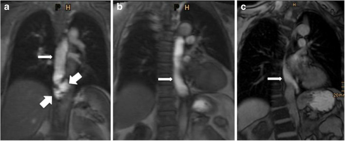
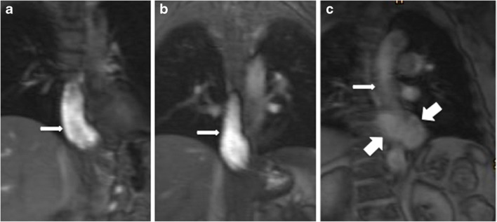
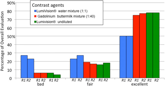
Methods: The protocol of this retrospective study was approved by the local Institutional Review Board. A hundred twenty-nine patients (146 examinations) underwent a dynamic MR swallowing study (at 1.5 T or 3.0 T) and received an oral contrast agent. Two readers evaluated the distention of the esophagus, contrast, and traceability of the bolus in a 3-point scale. A steady-state coherent sequence (B-FFE, TrueFISP) was used. The patients were divided into 3 different groups: 53 patients received gadolinium chelate (Dotarem®)-buttermilk mixture (GBM) in a dilution of 1:40 as an oral contrast agent; 44 patients received LumiVision® water mixture (LWM) in a dilution of 1:1 and 49 patients received LumiVision® (L) undiluted.
Results: GBM showed significantly better results in overall evaluation for both readers in contrast to LWM (p = .003, p = .002). L also reached significantly better results in overall evaluation than LWM in both readers (p = .004, p = .042). There was no significant difference in the overall evaluation between L and GBM (p = .914, p = .376).According to Landis and Koch, interobserver agreement was "substantial" (Cohen's kappa = 0.738) between both readers.
Conclusion: LumiVision® undiluted showed equal image quality compared to gadolinium-buttermilk mixture. The constellation of LumiVision® water mixture led to a clearly negative result in relation to the image quality compared to LumiVision® undiluted. Therefore, oral ingestion of LumiVision® undiluted is recommended for MR swallowing examinations.
Key points: • LumiVision® undiluted shows significantly better image quality in comparison to LumiVision® diluted in oral application in swallowing MRI. • LumiVision® undiluted shows equal image quality in comparison to gadolinium-buttermilk mixture in oral application. • Oral ingestion of LumiVision® undiluted can replace gadolinium-buttermilk mixture in oral MR examinations.
Keywords: Esophagus; Fundoplication, contrast agent; Magnetic resonance imaging.
© 2021. The Author(s).
Conflict of interest statement
The authors of this manuscript declare no relationships with any companies whose products or services may be related to the subject matter of the article.
Figures
References
MeSH terms
Substances
LinkOut - more resources
Full Text Sources
