

Giant intramuscular thigh lipoma: A case report and review of literature
- PMID: 33894509
- PMCID: PMC8091888
- DOI: 10.1016/j.ijscr.2021.105885
Giant intramuscular thigh lipoma: A case report and review of literature
Abstract
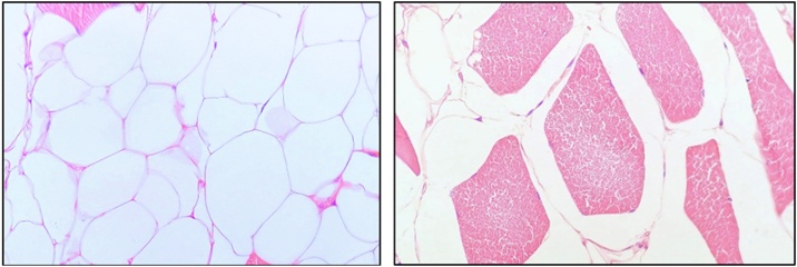
Introduction: Lipomas are the most common soft tissue tumor. Giant lipomas are defined by measuring at least 10 cm in diameter in one dimension or by a minimum of 1000 g. They often are asymptomatic; however, they can cause compression syndromes due to nerve damage and difficulties in walking.
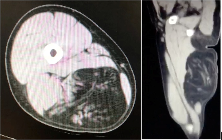
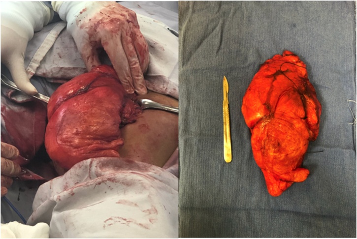
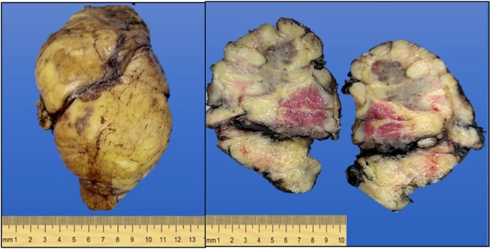
Presentation of case: We described the case of a 25-year-old female with no significant medical history who began her condition two years before her consultation. The patient referred to the appearance of a non-painful mass on her right thigh with progressive growth that hinders daily activities. A simple CT scan reported a 10.3 × 8.1 × 19.6 cm adipose mass with infiltration towards the semitendinosus muscle and the biceps femoris muscle. A free margin resection of the tumor was performed, and the involved muscles were preserved. The patient had a satisfactory postoperative outcome.
Discussion: Lipomas are common benign soft tissue tumors that arise from fatty tissue and may challenge surgical management due to their extension and dimensions; they often require delicate surgical intervention due to their potential risk of malignant transformation. We believe surgical pathologists and radiologists must draw attention to muscle involvement and the infiltrative pattern.
Conclusion: Giant lipomas should always raise awareness of malignant transformation. Radiological guidance should provide enough evidence to decide whether to do a biopsy or not; hence, saving the patient an extra invasive procedure. We recommend taking at least 1 cm of border margin while removing these tumors to avoid local recurrence.
Keywords: Lipoblasts; Lipocytes; Lipoma; Tumors.
Copyright © 2021 The Authors. Published by Elsevier Ltd.. All rights reserved.
Figures
References
-
- Zografos Gc, Kouerinis I., Kalliopi P., Karmen K., Evangelos M., Androulakis G. Vol. 109. 2002. Giant lipoma of the thigh in a patient with morbid obesity; pp. 1467–1468. (Plastic and Reconstructive Surgery). United States. - PubMed
-
- Bjerregaard P., Hagen K., Daugaard S., Kofoed H. Intramuscular lipoma of the lower limb. Long-term follow-up after local resection. J. Bone Joint Surg. Br. 1989;71(November (5)):812–815. - PubMed
-
- Davis C., Gruhn J. Giant lipoma of the thigh. Arch Surg. 1967;95(July (case 1)):1–6. - PubMed
-
- Phalen G.S., Kendrick J.I., Rodriguez J.M. Lipomas of the upper extremity. Am. J. Surg. 1971;121(3):298–306. - PubMed
LinkOut - more resources
Full Text Sources
Other Literature Sources
