. 2021 Jan-Jun:296:100701.
doi: 10.1016/j.jbc.2021.100701. Epub 2021 Apr 23.

Inhibition of acid sphingomyelinase by ambroxol prevents SARS-CoV-2 entry into epithelial cells

Affiliations

Inhibition of acid sphingomyelinase by ambroxol prevents SARS-CoV-2 entry into epithelial cells

Alexander Carpinteiro et al. J Biol Chem. 2021 Jan-Jun.

Abstract

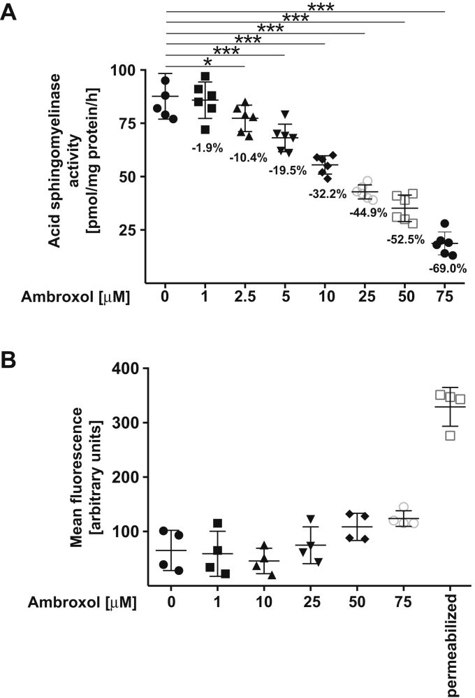
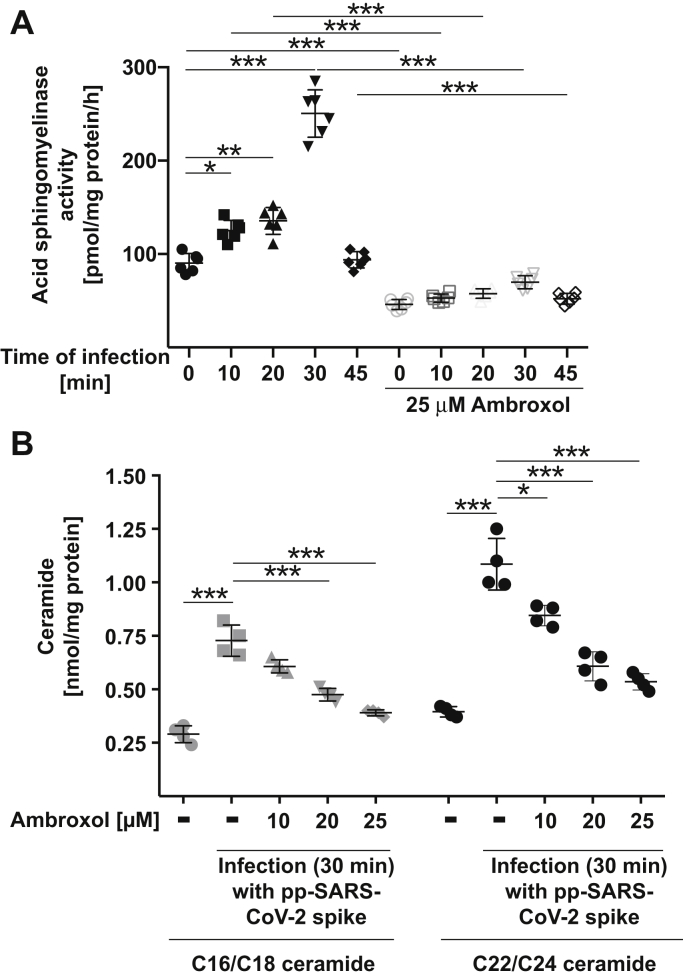
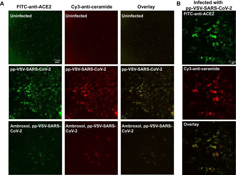
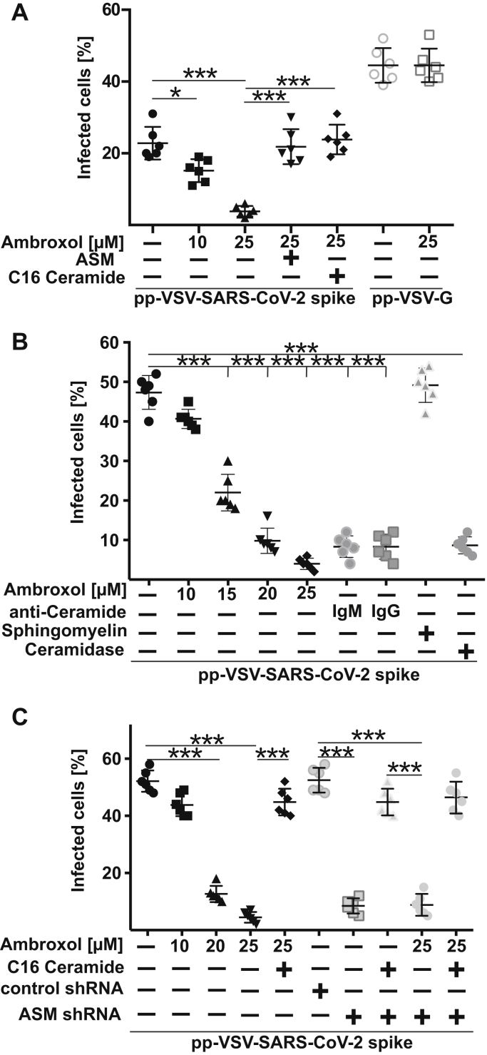
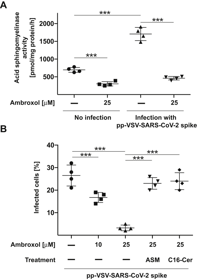
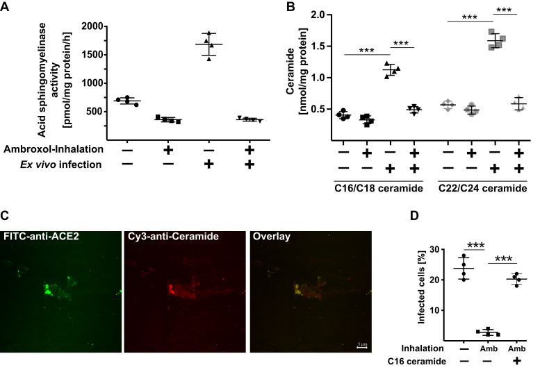
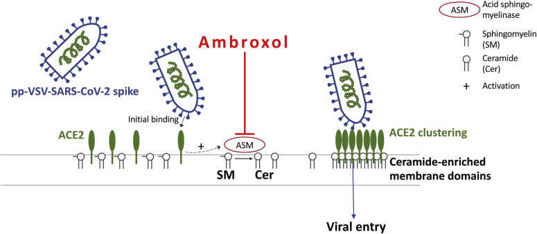
The acid sphingomyelinase/ceramide system has been shown to be important for cellular infection with at least some viruses, for instance, rhinovirus or severe acute respiratory syndrome coronavirus 2 (SARS-CoV-2). Functional inhibition of the acid sphingomyelinase using tricyclic antidepressants prevented infection of epithelial cells, for instance with SARS-CoV-2. The structure of ambroxol, that is, trans-4-[(2,4-dibromanilin-6-yl)-methyamino]-cyclohexanol, a mucolytic drug applied by inhalation, suggests that the drug might inhibit the acid sphingomyelinase and thereby infection with SARS-CoV-2. To test this, we used vesicular stomatitis virus pseudoviral particles presenting SARS-CoV-2 spike protein on their surface (pp-VSV-SARS-CoV-2 spike), a bona fide system for mimicking SARS-CoV-2 entry into cells. Viral uptake and formation of ceramide localization were determined by fluorescence microscopy, activity of the acid sphingomyelinase by consumption of [14C]sphingomyelin and ceramide was quantified by a kinase method. We found that entry of pp-VSV-SARS-CoV-2 spike required activation of acid sphingomyelinase and release of ceramide, events that were all prevented by pretreatment with ambroxol. We also obtained nasal epithelial cells from human volunteers prior to and after inhalation of ambroxol. Inhalation of ambroxol reduced acid sphingomyelinase activity in nasal epithelial cells and prevented pp-VSV-SARS-CoV-2 spike-induced acid sphingomyelinase activation, ceramide release, and entry of pp-VSV-SARS-CoV-2 spike ex vivo. The addition of purified acid sphingomyelinase or C16 ceramide restored entry of pp-VSV-SARS-CoV-2 spike into ambroxol-treated epithelial cells. We propose that ambroxol might be suitable for clinical studies to prevent coronavirus disease 2019.

Keywords: SARS-CoV-2; acid sphingomyelinase; ambroxol; ceramide; human nasal epithelial cells; infection.


Conflict of interest statement

Conflict of interest The authors declare that they have no conflicts of interest with the contents of this article.

Figures

Figure 1
Figure 1
Ambroxol reduces the activity of the acid sphingomyelinase.A, Vero-E6 cells were incubated with 1, 2.5, 5, 10, 25, 50, or 75 μM ambroxol or with solvent (0) for 60 min. Cells were lysed in 250 mM sodium acetate (pH 5.0) and 0.2% NP-40, and acid sphingomyelinase activity was determined by measuring the consumption of added [14C]sphingomyelin. The percent decrease of acid sphingomyelinase activity is given for easier quantification. Shown are the means ± SD of the acid sphingomyelinase activities from six independent experiments. ∗p < 0.05, ∗∗∗p < 0.001, ANOVA followed by post hoc Student's t tests. B, ambroxol did not exhibit cytotoxicity after treatment for 24 h. Cells were exposed for 24 h to 1, 10, 25, 50, or 75 μM ambroxol, and toxicity was measured by FITC–annexin V staining. FITC–annexin V staining was analyzed by flow cytometry. Permeabilized cells served as positive controls for the FITC–annexin V staining. Shown are means ± SD of the fluorescence (in arbitrary units) in the flow cytometry studies (n = 4); ANOVA, followed by post hoc Student's t tests. Results are presented in arbitrary units (a.u.). NP-40, Nonidet P40.
Figure 2
Figure 2
Ambroxol prevents activation of the acid sphingomyelinase and release of ceramide upon infection with pp-VSV-SARS-CoV-2 spike.A, Vero-E6 cells were preincubated with 25 μM ambroxol for 60 min or were left untreated. Cells were infected with pp-VSV-SARS-CoV-2 spike for the indicated times or were left uninfected (0). Cells were lysed in 250 mM sodium acetate (pH 5.0) and 0.2% NP-40, and acid sphingomyelinase activity was determined by measuring the consumption of added [14C]sphingomyelin. Displayed are the means ± SD of the activity of the acid sphingomyelinase from six independent experiments. ∗p < 0.05, ∗∗p < 0.01, ∗∗∗p < 0.001, ANOVA followed by post hoc Student's t tests. Closed symbols give the time course without ambroxol, and open symbols give the time course with 25 μM ambroxol. B, cells were treated with 10, 20, or 25 μM ambroxol for 1 h and infected with pp-SARS-CoV-2 spike for 30 min or left untreated or uninfected, washed, and organically extracted to quantify C16/C18 ceramide and C22/C24 ceramide levels by using the ceramide kinase method. Displayed are the means ± SD of the ceramide concentrations from each six independent experiments. ∗p < 0.05, ∗∗∗p < 0.001, ANOVA, followed by post hoc Student's t tests, as indicated or compared with the corresponding value without inhibitor. Gray symbols represent 16/C18 ceramides, and black symbols represent C22/24 ceramides. NP-40, Nonidet P40; pp-VSV-SARS-CoV-2, vesicular stomatitis virus pseudoviral particles presenting SARS-CoV-2 spike protein on their surface.
Figure 3
Figure 3
ACE2 clusters in ceramide-enriched membrane domains upon infection.A and B, infection of Vero-E6 cells with pp-VSV-SARS-CoV-2 results in formation of ceramide-enriched membrane domains that cluster ACE2, which is prevented by treatment with ambroxol. Cells were infected for 30 min, fixed, stained with Cy3-coupled anti-ceramide and FITC–anti-ACE2 antibodies, and analyzed by confocal microscopy. A, shows a low magnification overview to visualize the abundance of ceramide-enriched membrane domains clustering ACE2. B, displays a higher magnification of infected cells. Please note that infection does not only induces ceramide but also increases surface expression of ACE2 (14). Shown is a representative result from six independent experiments. ACE2, angiotensin-converting enzyme 2; pp-VSV-SARS-CoV-2, vesicular stomatitis virus pseudoviral particles presenting SARS-CoV-2 spike protein on their surface.
Figure 4
Figure 4
Ambroxol prevents infection with SARS-CoV-2.A, Vero-E6 cells were pretreated with 10 μM or 25 μM ambroxol for 1 h or left untreated and were infected with pp-VSV-SARS-CoV-2 spike or pp-VSV-G for 24 h. Infection was measured by the expression of eGFP in the cells. Ambroxol blocked the infection of the cells with pp-VSV-SARS-CoV-2 spike (closed symbols) but had no influence on infection with pp-VSV-G (open symbols). Reconstitution of ceramide in Vero-E6 cells that had been treated with 25 μM ambroxol by the addition of 10 μM C16 ceramide or 0.2 U/ml acid sphingomyelinase (ASM) restored infection of the cells with pp-VSV-SARS-CoV-2 spike. Shown are the means ± SD of the percentage of infected cells from six independent experiments. ∗∗∗p< 0.001, ANOVA, followed by post hoc Student's t tests. B, higher concentrations of the pp-VSV-SARS-CoV-2 spike result in a higher percentage of infected cells but do not alter the ability of ambroxol to dose dependently inhibit infection of Vero-E6 cells. Neutralization of surface ceramide with two different anti-ceramide antibodies, that is, a monoclonal IgM antibody and an IgG antibody, or treatment with a ceramidase also prevented infection with pp-VSV-SARS-CoV-2 spike. Addition of sphingomyelin (50 μM) was without effect on the infection. Given are the means ± SD of the percentage of infected cells from six independent experiments. ∗∗∗p< 0.001, ANOVA, followed by post hoc Student's t tests. Black symbols are untreated and ambroxol-treated cells, and gray symbols represent cells constituted with anti-ceramide antibodies or sphingomyelin. C, treatment of Caco-2 cells with ambroxol dose dependently prevented infection with pp-VSV-SARS-CoV-2 spike. Likewise, transfection of Caco-2 cells with shRNA targeting the acid sphingomyelinase prevented infection, whereas a control shRNA had no effect on the infection. Addition of ambroxol to cells transfected with acid sphingomyelinase targeting shRNA had no additional effects. Displayed are the means ± SD of the percentage of infected cells from six independent experiments. ∗∗∗p< 0.001, ANOVA, followed by post hoc Student's t tests. Closed symbols are untransfected cells, and open symbols represent transfected cells. eGFP, enhanced GFP; pp-VSV-SARS-CoV-2, vesicular stomatitis virus pseudoviral particles presenting SARS-CoV-2 spike protein on their surface; SARS-CoV-2, severe acute respiratory syndrome coronavirus 2.
Figure 5
Figure 5
Ambroxol prevents activation of the acid sphingomyelinase by an infection of freshly isolated human nasal epithelial cells with pp-VSV-SARS-Cov-2 spike in vitro.A, freshly isolated human nasal epithelial cells were incubated with 25 μM ambroxol for 1 h or left untreated and infected with pseudoviral particles of pp-VSV-SARS-CoV-2 spike for 30 min or left uninfected. The medium was removed, cells were lysed, and activity of the acid sphingomyelinase was determined by consumption of added [14C]sphingomyelin. Given are the means ± SD of the acid sphingomyelinase activity from four independent experiments. ∗∗∗p < 0.001, ANOVA, followed by post hoc Student's t tests. B, freshly isolated human nasal epithelial cells were incubated with 10 μM or 25 μM ambroxol for 1 h and infected with pp-VSV-SARS-CoV-2 spike. Infection was determined by counting eGFP-positive cells in at least 500 cells/sample. Reconstitution of ceramide by application of 0.2 U/ml purified acid sphingomyelinase, or 10 μM C16 ceramide (C16-Cer) restored infection in human nasal epithelial cells treated with ambroxol. Given are the means ± SD of the percentage of eGFP-positive, that is, infected cells, from four independent experiments. ∗∗∗p < 0.001, ANOVA followed by post hoc Student's t tests. eGFP, enhanced GFP; pp-VSV-SARS-Cov-2, vesicular stomatitis virus pseudoviral particles presenting SARS-CoV-2 spike protein on their surface.
Figure 6
Figure 6
In vivo administration of ambroxol prevents infection of nasal epithelial cells with pp-VSV-SARS-CoV-2 spike ex vivo. Nasal epithelial cells were obtained from volunteers. The volunteers then inhaled 2 ml ambroxol, that is, 15 mg ambroxol. Nasal epithelial cells were obtained from the opposite nasal cavity 1 h after inhalation. All cells were immediately washed after isolation, and the cell pellets were resuspended in cell culture medium and infected with pp-VSV-SARS-CoV-2 spike or left uninfected for 30 min (AC) to determine acid sphingomyelinase (A), ceramide (B), and ceramide-enriched membrane domains with ACE2 clusters (C) or for 1 h (D) to determine infection (D). A, to determine acid sphingomyelinase activity, cell pellets were lysed in 50 mM sodium acetate (pH 5.0) and 0.2% NP-40, and the activity of the acid sphingomyelinase was determined by measuring the consumption of added [14C]sphingomyelin. B, cells were organically extracted, and ceramide was determined by the ceramide kinase assay method. Black symbols indicate C16/C18 ceramides, and gray symbols indicate C22/C24 ceramides. C, freshly isolated nasal epithelial cells were infected with pp-VSV-SARS-CoV-2 for 30 min, washed, fixed in 1% PFA for 10 min, washed and stained with Cy3-coupled anti-ceramide and FITC–anti-ACE2 antibodies. Shown are representative results from four independent experiments. D, cells were infected with pp-VSV-SARS-CoV-2 spike for 1 h, washed, and expression of eGFP was determined after 24 h in at least 500 epithelial cells per sample in randomly chosen microscopic fields. If indicated, C16 ceramide (10 μM) was reconstituted during the infection. A, B, and D show the means ± SD from four volunteers. ∗∗∗p < 0.001, ANOVA, followed by post hoc Student's t tests. ACE2, angiotensin-converting enzyme 2; Amb, ambroxol inhalation; eGFP, enhanced GFP; NP-40, Nonidet P40; PFA, paraformaldehyde; pp-VSV-SARS-CoV-2.
Figure 7
Figure 7
Scheme of the proposed inhibition of pp-SARS-CoV-2 spike infections by ambroxol. Initial binding of pp-VSV-SARS-CoV-2 spike results in activation of the acid sphingomyelinase, formation of surface ceramide, and formation of ceramide-enriched membrane platforms that serve to trap and cluster activated ACE2 receptors. The clustering of ACE2 in ceramide-enriched membrane platforms might facilitate signaling via this receptor finally resulting in internalization but also brings the virus into close proximity to proteases required for uptake. ACE2, angiotensin-converting enzyme 2; pp-VSV-SARS-CoV-2, vesicular stomatitis virus pseudoviral particles presenting SARS-CoV-2 spike protein on their surface.

References

    1. Zhou P., Yang X.L., Wang X.G., Hu B., Zhang L., Zhang W., Si H.R., Zhu Y., Li B., Huang C.L., Chen H.D., Chen J., Luo Y., Guo H., Jiang R.D. A pneumonia outbreak associated with a new coronavirus of probable bat origin. Nature. 2020;579:270–273. - PMC - PubMed
    1. Serafim R.B., Povoa P., Souza-Dantas V., Andre C., Kalil A.C., Salluh J.I.F. Clinical course and outcomes of critically ill patients with COVID-19 infection: A systematic review. Clin. Microbiol. Infect. 2021;27:47–54. - PMC - PubMed
    1. Wrapp D., Wang N., Corbett K.S., Goldsmith J.A., Hsieh C.L., Abiona O., Graham B.S., McLellan J.S. Cryo-EM structure of the 2019-nCoV spike in the prefusion conformation. Science. 2020;367:1260–1263. - PMC - PubMed
    1. Lan J., Ge J., Yu J., Shan S., Zhou H., Fan S., Zhang Q., Shi X., Wang Q., Zhang L., Wang X. Structure of the SARS-CoV-2 spike receptor-binding domain bound to the ACE2 receptor. Nature. 2020;581:215–220. - PubMed
    1. Wang Q., Zhang Y., Wu L., Niu S., Song C., Zhang Z., Lu G., Qiao C., Hu Y., Yuen K.Y., Wang Q., Zhou H., Yan J., Qi J. Structural and functional basis of SARS-CoV-2 entry by using human ACE2. Cell. 2020;181:894–904. - PMC - PubMed

Publication types

MeSH terms

LinkOut - more resources