. 2021 Jul 10:510:67-78.
doi: 10.1016/j.canlet.2021.04.011. Epub 2021 Apr 22.

The TIM3/Gal9 signaling pathway: An emerging target for cancer immunotherapy

Affiliations

The TIM3/Gal9 signaling pathway: An emerging target for cancer immunotherapy

Sashi Kandel et al. Cancer Lett. .

Abstract

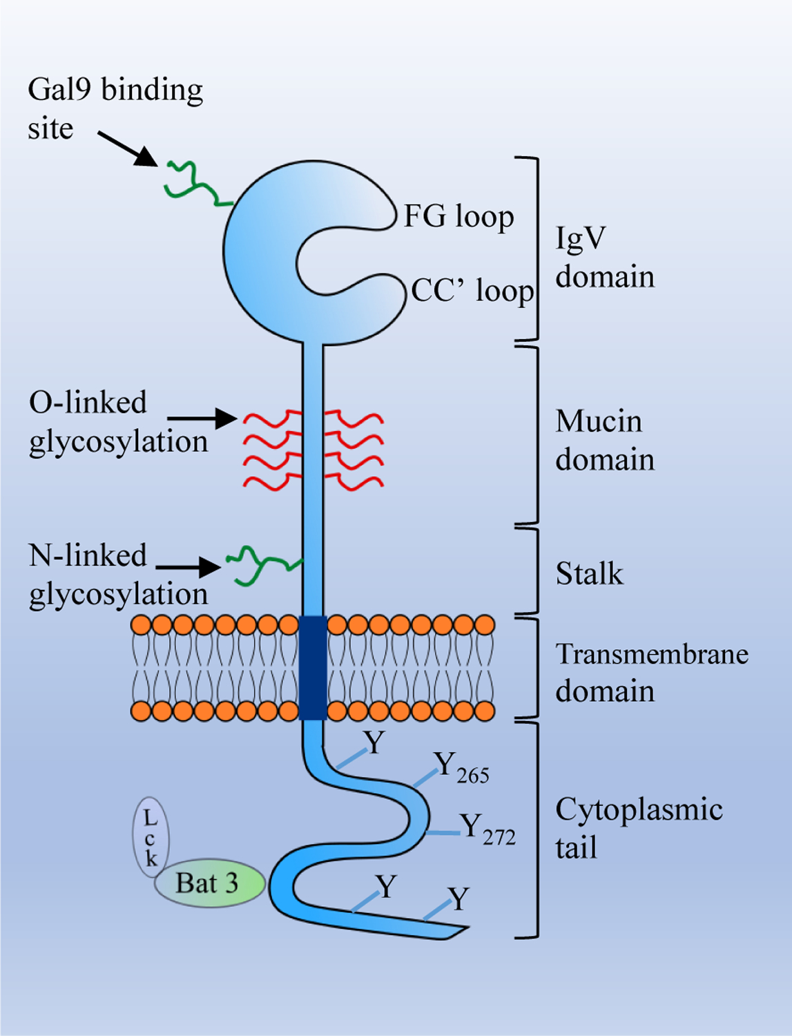
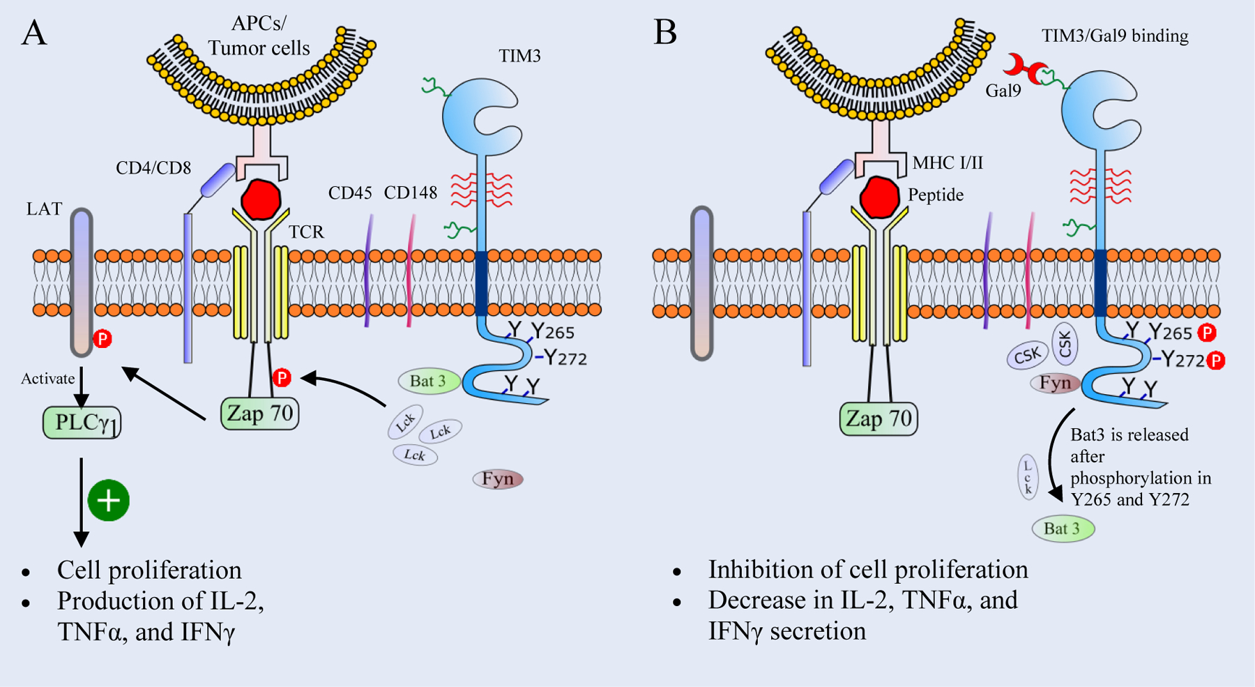
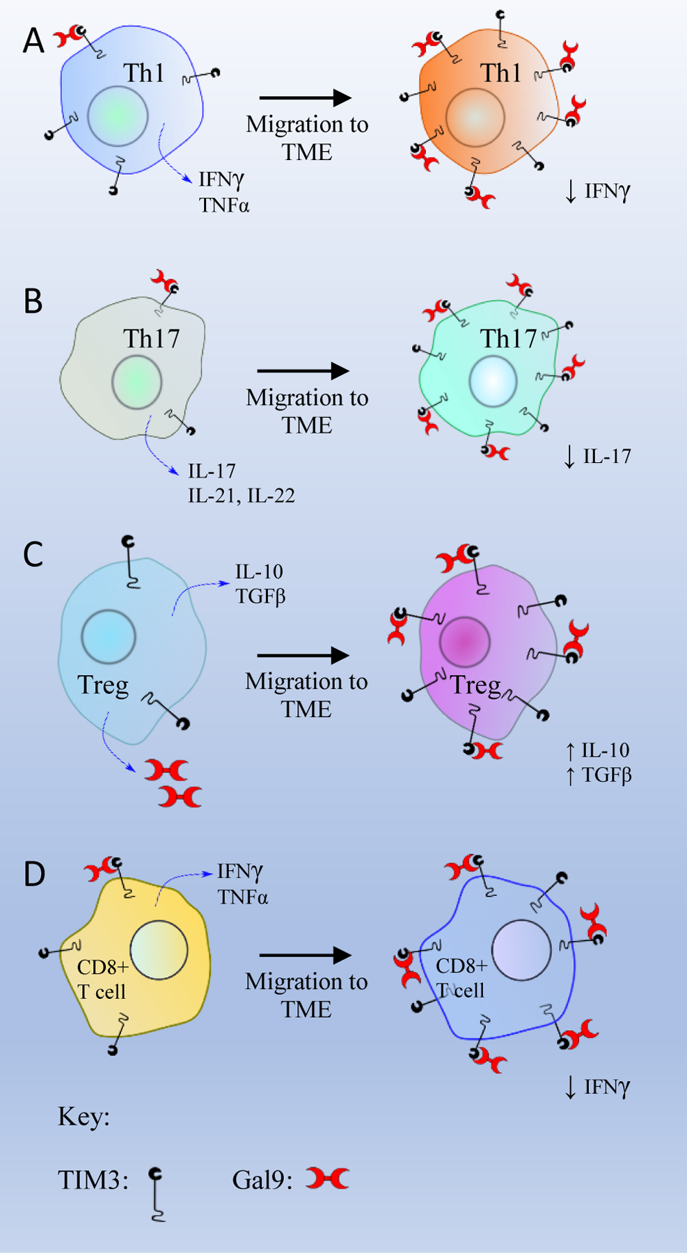
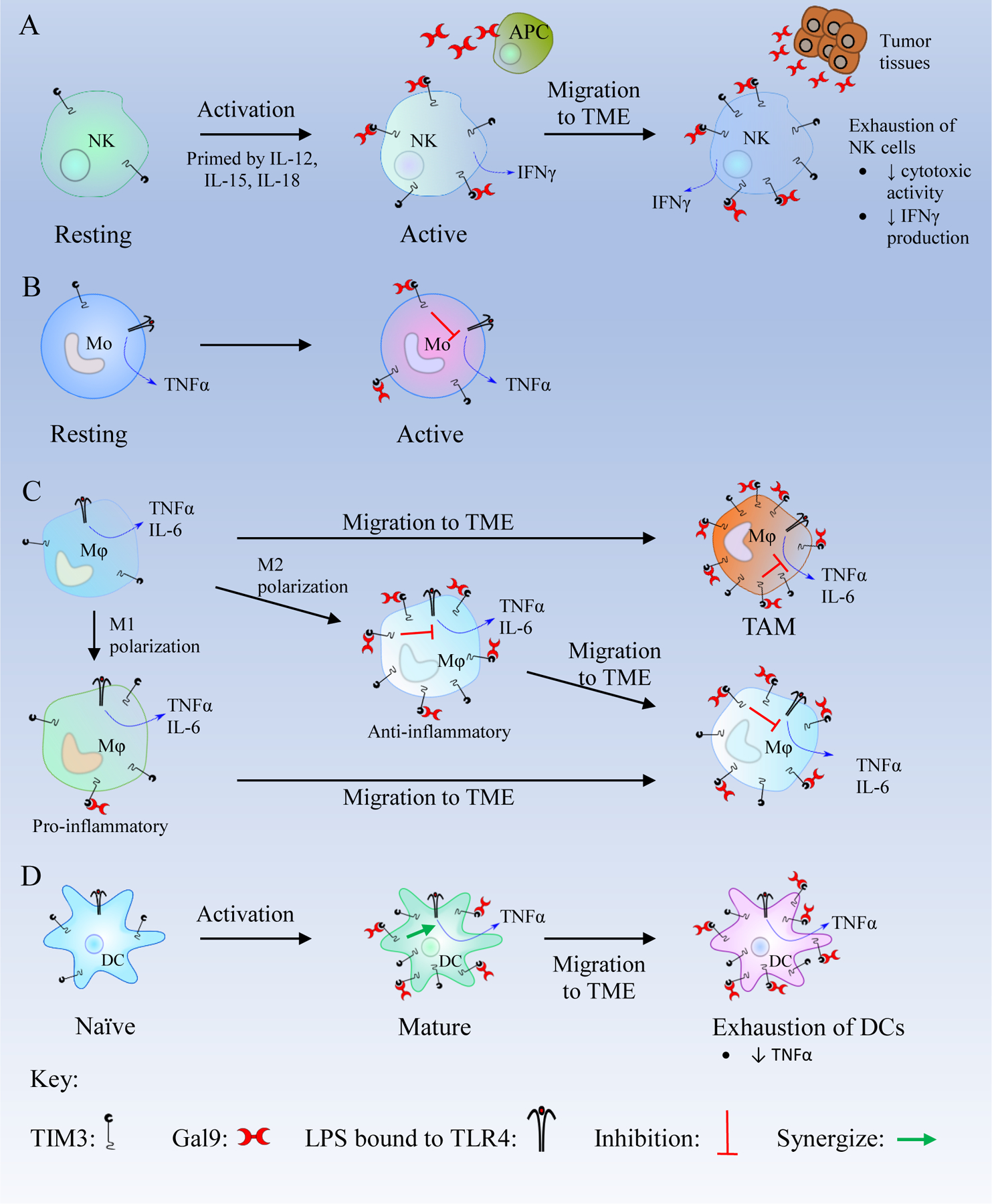
Immune checkpoint blockade has shown unprecedented and durable clinical response in a wide range of cancers. T cell immunoglobulin and mucin domain 3 (TIM3) is an inhibitory checkpoint protein that is highly expressed in tumor-infiltrating lymphocytes. In various cancers, the interaction of TIM3 and Galectin 9 (Gal9) suppresses anti-tumor immunity mediated by innate as well as adaptive immune cells. Thus, the blockade of the TIM3/Gal9 interaction is a promising therapeutic approach for cancer therapy. In addition, co-blockade of the TIM3/Gal9 pathway along with the PD-1/PD-L1 pathway increases the therapeutic efficacy by overcoming non-redundant immune resistance induced by each checkpoint. Here, we summarize the physiological roles of the TIM3/Gal9 pathway in adaptive and innate immune systems. We highlight the recent clinical and preclinical studies showing the involvement of the TIM3/Gal9 pathway in various solid and blood cancers. In addition, we discuss the potential of using TIM3 and Gal9 as prognostic and predictive biomarkers in different cancers. An in-depth mechanistic understanding of the blockade of the TIM3/Gal9 signaling pathway in cancer could help in identifying patients who respond to this therapy as well as designing combination therapies.

Keywords: Immune checkpoint blockade; Immune checkpoint receptors; Immune suppression; Immuno-oncology; TIM3/Gal9.


Conflict of interest statement

Conflict of Interest

The authors declare that they have no competing interests.

Declaration of interests

⊠The authors declare that they have no known competing financial interests or personal relationships that could have appeared to influence the work reported in this paper.

Figures

Figure 1.
Figure 1.. The structure of TIM3.
The extracellular region of T cell immunoglobulin and mucin domain-containing protein 3 (TIM3) consists of an IgV domain, a mucin domain, and a stalk domain. The transmembrane region consists of a transmembrane domain and the intracellular region consists of a cytoplasmic tail with five tyrosine residues. The IgV domain contains binding sites for its ligands. Phosphatidylserine (Ptdser), carcinoembryonic antigen-related cell adhesion molecule (CEACAM1), and high mobility group box 1 (HMGB1) bind to the FG-CC’ loop, while Gal9 binds to the N-linked glycan.
Figure 2.
Figure 2.. Schematics of the TIM3/Gal9 signaling pathway in T cells.
(A) In an unbound state, the cytoplasmic tail of T cell immunoglobulin and mucin domain-containing protein 3 (TIM3) binds to HLA-B associated transcript 3 (Bat3) which helps to recruit the tyrosine kinase Lck. Lck clusters then phosphorylate the CD3 domain of TCR, leading to the recruitment of the tyrosine kinase Zap 70. Zap 70 then phosphorylates linker adaptor protein (LAT), which activates the phospholipase Cγ1 (PLCγ1) to produce second messengers like Inositol trisphosphate (IP3) and diacylglycerol (DAG), leading to the production of cytokines and T cell proliferation. (B) Upon the TIM3/Gal9 interaction, Y265 and Y272 in the cytoplasmic tail of TIM3 are phosphorylated, resulting in the release of Bat3 and Lck. The tyrosine kinase Fyn is recruited to the cytoplasmic tail of TIM3 and helps in the clustering of C-terminal c-Src kinase (Csk). Csk clusters promote phosphorylation of the C-terminal tyrosine of Lck and inhibit its catalytic activity. Subsequently this leads to the inhibition of T cell proliferation and suppression of the production of IL-2, TNFα, and IFNγ.
Figure 3:
Figure 3:. The role of the TIM3/Gal9 interaction on different T cell subsets.
(A) T helper 1 cell (Th1), (B) T helper 17 cell (Th17), (C) T regulatory cells (Tregs), and (D) CD8+ T cell.
Figure 4.
Figure 4.. Schematics of the TIM3/Gal9 pathway in innate immune cells.
(A) Resting natural killer (NK) cells become active in the presence of IL-12, IL-15, and IL-18 and express more T cell immunoglobulin and mucin-containing protein 3 (TIM3). In active NK cells, TIM3/Galectin 9 (Gal9) binding enhances interferon γ (IFNγ) secretion. After migration to the tumor microenvironment (TME), NK cells are exposed to high levels of Gal9 which leads to their exhaustion. (B) Active monocytes (Mo) express a high number of TIM3 receptors. The TIM3/Gal9 interaction inhibits the Lipopolysaccharide (LPS)/Toll like receptor 4 (TLR4) pathway and decreases the secretion of TNFα. (C) Macrophages (Mφ) in the TME overexpress TIM3. Macrophages are polarized to M1 and M2 phenotypes. M1 macrophages are pro-inflammatory and express a low level of TIM3. M1 macrophages can migrate to the TME and may transform to M2 macrophages. M2 macrophages are anti-inflammatory and express high levels of TIM3. TIM3/Gal9 binding in M2 macrophages inhibits LPS/TLR4 pathway and decreases the production of inflammatory cytokines like Tumor Necrosis Factor α (TNFα), interleukin-6 (IL-6), and IL-12. After migrating to the TME, M2 macrophages overexpress TIM3, and the TIM3/Gal9 binding leads to the inhibition of the LPS/TLR4 pathway to suppress the immune response. (D) Mature DCs express a high level of TIM3, and the TIM3/Gal9 binding synergizes with the LPS/TLR4 pathway which produces TNFα. In Dendritic cells (DCs) within the TME, the TIM3/Gal9 interaction suppresses CXCL9 expression, thus inhibiting innate immunity and promote tumor growth.

References

    1. Darvin P, Toor SM, Sasidharan Nair V, Elkord E, Immune checkpoint inhibitors: recent progress and potential biomarkers, Exp Mol Med, 50 (2018) 1–11. - PMC - PubMed
    1. Pardoll DM, The blockade of immune checkpoints in cancer immunotherapy, Nat Rev Cancer, 12 (2012) 252–264. - PMC - PubMed
    1. Zhu C, Anderson AC, Schubart A, Xiong H, Imitola J, Khoury SJ, Zheng XX, Strom TB, Kuchroo VK, The Tim-3 ligand galectin-9 negatively regulates T helper type 1 immunity, Nat Immunol, 6 (2005) 1245–1252. - PubMed
    1. Freeman GJ, Casasnovas JM, Umetsu DT, DeKruyff RH, TIM genes: a family of cell surface phosphatidylserine receptors that regulate innate and adaptive immunity, Immunol Rev, 235 (2010) 172–189. - PMC - PubMed
    1. Chiba S, Baghdadi M, Akiba H, Yoshiyama H, Kinoshita I, Dosaka-Akita H, Fujioka Y, Ohba Y, Gorman JV, Colgan JD, Hirashima M, Uede T, Takaoka A, Yagita H, Jinushi M, Tumor-infiltrating DCs suppress nucleic acid-mediated innate immune responses through interactions between the receptor TIM-3 and the alarmin HMGB1, Nat Immunol, 13 (2012) 832–842. - PMC - PubMed

Publication types

Substances