. 2021 Apr 24;6(1):169.
doi: 10.1038/s41392-021-00591-7.

The olfactory route is a potential way for SARS-CoV-2 to invade the central nervous system of rhesus monkeys

Affiliations

The olfactory route is a potential way for SARS-CoV-2 to invade the central nervous system of rhesus monkeys

Li Jiao et al. Signal Transduct Target Ther. .

Abstract

Neurological manifestations are frequently reported in the COVID-19 patients. Neuromechanism of SARS-CoV-2 remains to be elucidated. In this study, we explored the mechanisms of SARS-CoV-2 neurotropism via our established non-human primate model of COVID-19. In rhesus monkey, SARS-CoV-2 invades the CNS primarily via the olfactory bulb. Thereafter, viruses rapidly spread to functional areas of the central nervous system, such as hippocampus, thalamus, and medulla oblongata. The infection of SARS-CoV-2 induces the inflammation possibly by targeting neurons, microglia, and astrocytes in the CNS. Consistently, SARS-CoV-2 infects neuro-derived SK-N-SH, glial-derived U251, and brain microvascular endothelial cells in vitro. To our knowledge, this is the first experimental evidence of SARS-CoV-2 neuroinvasion in the NHP model, which provides important insights into the CNS-related pathogenesis of SARS-CoV-2.


Conflict of interest statement

The authors declare no competing interests.

Figures
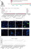
Fig. 1
Fig. 1
SARS-CoV-2 invades the CNS in rhesus monkeys post intranasal inoculation. a Scheme of the experimental design of intranasal SARS-CoV-2 inoculation. Five rhesus monkeys (3–5 years old) were intranasally inoculated with 1 × 107 PFU of SARS-CoV-2 in 1 mL PBS. Monkeys were observed for clinical signs, including body temperature, body weight, sample collection, chest radiograph, and necropsies as indicated. b Viral load in CNS tissues from SARS-CoV-2-inoculated rhesus macaques. On 1, 4, 7, and 14 dpi, animals were necropsied and tissue samples in the CNS were collected. Viral genomic RNA was quantitated by quantitative real-time polymerase chain reaction (qRT-PCR). c Immunofluorescence (IF) staining of viral N protein (green) in CNS tissues from rhesus macaques infected with SARS-CoV-2. The sections were counterstained with DAPI for nuclei. Each data point represents an independent field of view from slides subjected to IF. Scale bar, 50 μm. d qRT-PCR analysis of angiotensin-converting enzyme 2 (ACE2) and TMPRSS2 mRNA expression in the brain tissues from the uninfected rhesus macaque
Fig. 2
Fig. 2
SARS-CoV-2 invasion results in inflammation and pathological changes in the CNS. a Hematoxylin and eosin (H&E) staining showed that intranasal SARS-CoV-2 inoculation caused different degrees of neuronal death, glial hyperplasia, and edema and perivascular inflammatory cell infiltration in the brain compared with PBS control (0 dpi), which is described in the text. Scale bar (upper panel), 50 μm; scale bar (lower panel), 20 μm.(b) Immunochemistry (IHC) analysis of CD68-positive cells in the brains of rhesus macaques on 0, 1, 4, 7 and 14 dpi. Each data point represents an independent field of view from slides subjected to IHC. Arrow, positive staining. Scale bar, 100 μm. c Concentrations of inflammatory cytokines in the brains of rhesus macaques infected with SARS-CoV-2. d Concentrations of inflammatory cytokines in the serum of rhesus macaques infected with SARS-CoV-2
Fig. 3
Fig. 3
Intracranial inoculation with SARS-CoV-2 causes the neuroinflammation in multiple brain regions of rhesus monkeys. Rhesus monkeys were intracranially inoculated with a high (MM-C-H) and low (MM-C-L) dose of SARS-CoV-2. On 9 dpi, 3 animals (including PBS Control) were necropsied for the following analyses. a Viral genomic RNA in the CNS tissues (MM-C-H-L) and CSF was quantitated by qRT-PCR. b IF staining of N protein in the CNS tissues. Scale bar, 100 μm. The images in the solid line boxes are zoomed in from those in the dotted line boxes. c IF double staining for viral N protein (red) and cell surface markers (green) in thalamic infected with a high dose of SARS-CoV-2. Scale bar, 100 μm. d H&E staining in the brains of rhesus macaques intracranially inoculated with a high (MM-C-H) and low (MM-C-L) dose of SARS-CoV-2 or PBS. Scale bar (left panel), 50 μm; scale bar (right panel), 20 μm. e IHC for CD68-positive cells in the brains of rhesus macaques infected with a high dose of SARS-CoV-2 (MM-C-H) or PBS. Scale bar, 100 μm. f Concentrations of inflammatory cytokines in the brains of rhesus macaques. g Concentrations of inflammatory cytokines in the serum of rhesus macaques
Fig. 4
Fig. 4
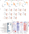
CNS cells in hippocampus undergo a hyperbiosynthetic, hypermetabolic, and mitochondrial disorders in response to SARS-CoV-2 infections. a t-SNE projection of cells from single-cell sequencing. b Heatmap of receptor genes for SARS-CoV-2 in the hippocampus of rhesus macaques. c Enriched GO terms in SARS-CoV-2-infected hippocampus and SARS-CoV-2 compared with control. d Heatmap of differentially expressed genes (DEGs) from the hippocampus infected with SARS-CoV-2 compared with control

References

    1. Wang C, Horby PW, Hayden FG, Gao GF. A novel coronavirus outbreak of global health concern. Lancet. 2020;395:470–473. - PMC - PubMed
    1. Morens DM, Daszak P, Taubenberger JK. Escaping Pandora’s box - another novel coronavirus. N. Engl. J. Med. 2020;382:1293–1295. doi: 10.1056/NEJMp2002106. - DOI - PubMed
    1. Moriguchi T, et al. A first case of meningitis/encephalitis associated with SARS-coronavirus-2. Int. J. Infect. Dis. 2020;94:55–58. doi: 10.1016/j.ijid.2020.03.062. - DOI - PMC - PubMed
    1. Mao L, et al. Neurologic manifestations of hospitalized patients with coronavirus disease 2019 in Wuhan, China. JAMA Neurol. 2020;77:683–690. doi: 10.1001/jamaneurol.2020.1127. - DOI - PMC - PubMed
    1. Asadi-Pooya AA, Simani L. Central nervous system manifestations of COVID-19: a systematic review. J. Neurol. Sci. 2020;413:116832. doi: 10.1016/j.jns.2020.116832. - DOI - PMC - PubMed

Publication types

MeSH terms