. 2021 Apr;33(4):042004.
doi: 10.1063/5.0048123. Epub 2021 Apr 6.

Effect of self-assembly on fluorescence in magnetic multiphase flows and its application on the novel detection for COVID-19

Affiliations

Effect of self-assembly on fluorescence in magnetic multiphase flows and its application on the novel detection for COVID-19

Xiang Li et al. Phys Fluids (1994). 2021 Apr.

Abstract

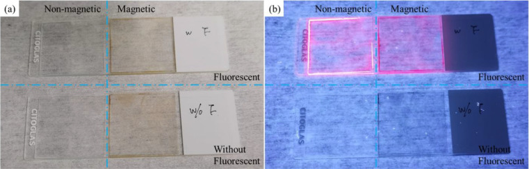
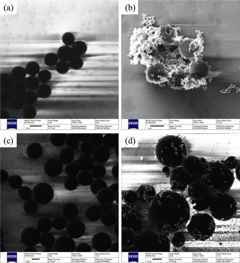
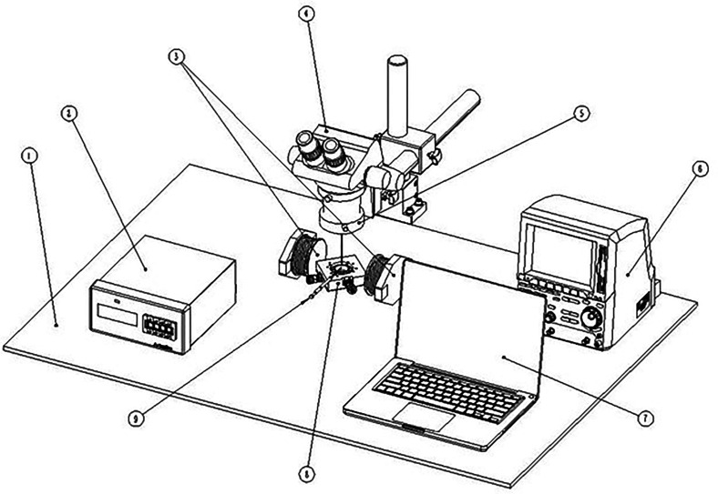
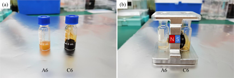
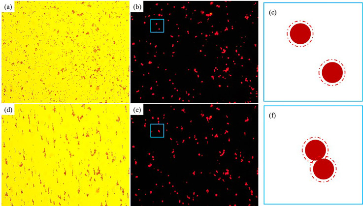
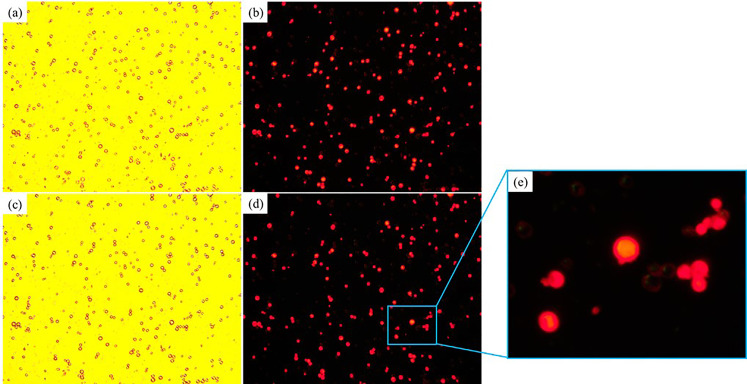
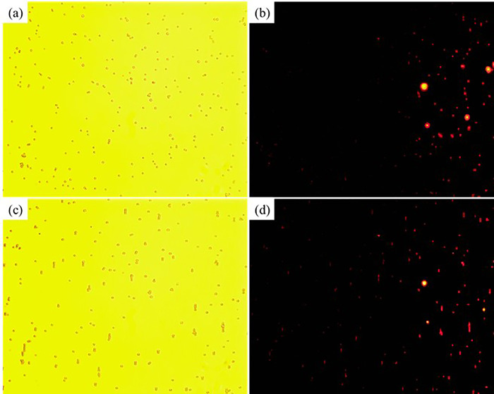
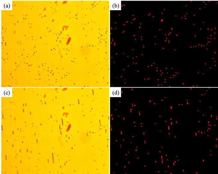
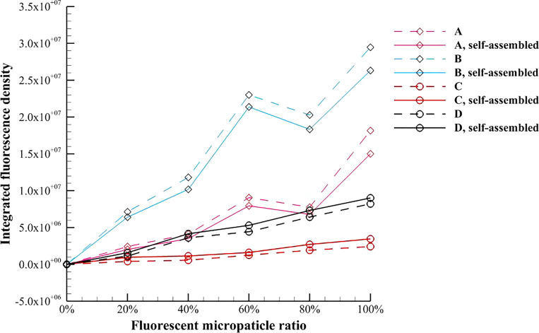
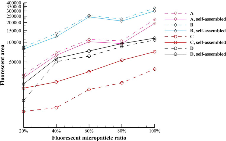
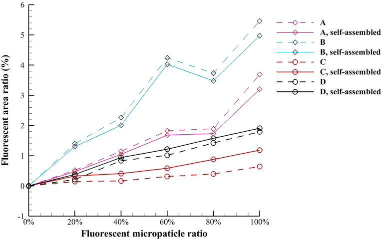
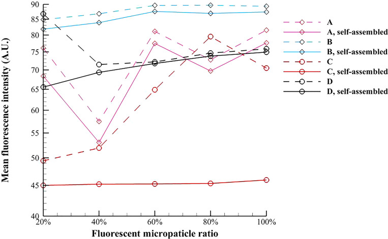
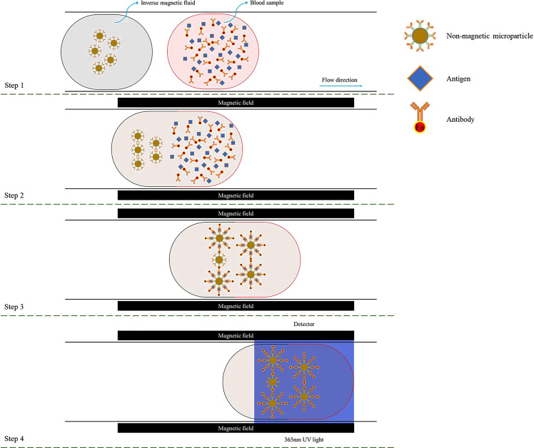
In the present study, the magnetic field induced self-assembly processes of magnetic microparticles in an aqueous liquid (the pure magnetic fluid) and nonmagnetic microparticles in ferrofluid (the inverse magnetic fluid) are experimentally investigated. The microparticles are formed into chain-like microstructures in both the pure magnetic fluid and the inverse magnetic fluid by applying the external magnetic field. The fluorescence parameters of these self-assembled chain-like microstructures are measured and compared to those without the effect of magnetic field. It is found that the fluorescence in the pure magnetic fluid is weakened, because the scattering and illuminating areas are reduced in the microstructures. On the contrary, the fluorescence in the inverse magnetic fluid is enhanced, because more fluorescent nonmagnetic microparticles are enriched and become detectable under the effect of the magnetic dipole force and the magnetic levitational force, and their unnecessary scattering can be absorbed by the surrounding ferrofluid. The average enhancement of the fluorescence area ratio in the inverse magnetic fluid with 3 μm nonmagnetic microparticles reaches 112.92%. The present work shows that the inverse magnetic fluid has advantages such as low cost, no scattering effect, stable fluorescence intensity, and relatively low magnetic resistance. In the end, a prototype design for the novel detection of coronavirus disease 2019 based on the magnetic field induced self-assembly in the inverse magnetic fluid is proposed, which could support the epidemic prevention and control.


Figures

FIG. 1.
FIG. 1.
Comparison for the fluorescent responses of the 3 μm nonmagnetic microparticles, the 3 μm nonmagnetic fluorescent microparticles, the 3 μm magnetic microparticles, and the 3 μm magnetic fluorescent microparticles on glass slides: (a) white light; (b) 365 nm ultraviolet light.
FIG. 2.
FIG. 2.
The surface topographies of the microparticles obtained by helium ion microscopy: (a) 3 μm nonmagnetic microparticles, (b) 3 μm nonmagnetic fluorescent microparticles, (c) 5 μm magnetic microparticles, and (d) 5 μm magnetic fluorescent microparticles.
FIG. 3.
FIG. 3.
Experimental setup to observe the self-assembly process in the antibody detection under an external uniform magnetic field. (① The workbench, ② the monitor of Tesla meter, ③ a couple of electromagnets, ④ the electronic ocular of microscope (i.e., CCD camera), ⑤ the digital metallographic microscope, ⑥ the precision programable DC power supply, ⑦ the laptop, ⑧ the objective table with light source [365 nm ultraviolet light] and condenser lens, and ⑨ the probe of Tesla meter.).
FIG. 4.
FIG. 4.
Variation of the magnetic intensity H with the current of DC power supply A.
FIG. 5.
FIG. 5.
Comparison of the pure magnetic fluid and the inverse magnetic fluid used in the present work: (a) without magnet; (b) with magnet. The north and south poles of the magnet are indicated by the letters N and S.
FIG. 6.
FIG. 6.
The magnetic field induced self-assembly process of 3 μm magnetic microparticles in the A06 test magnetic multiphase fluid: (a) white light, without magnetic field; (b) 365 nm ultraviolet light, without magnetic field; (c) schematic diagram, without magnetic field; (d) white light, with magnetic field; (e) 365 nm ultraviolet light, with magnetic field; (f) schematic diagram, with magnetic field.
FIG. 7.
FIG. 7.
The magnetic field induced self-assembly process of 5 μm magnetic microparticles in the B06 test magnetic multiphase fluid: (a) white light, without magnetic field; (b) 365 nm ultraviolet light, without magnetic field; (c) white light, with magnetic field; (d) 365 nm ultraviolet light, with magnetic field; (e) partial enlarged detail, with magnetic field.
FIG. 8.
FIG. 8.
The magnetic field induced self-assembly process of 3 μm nonmagnetic microparticles in the C06 test magnetic multiphase fluid: (a) white light, without magnetic field; (b) 365 nm ultraviolet light, without magnetic field; (c) schematic diagram, with magnetic field; (d) white light, with magnetic field.
FIG. 9.
FIG. 9.
The magnetic field induced self-assembly process of 5 μm magnetic microparticles in the D06 test magnetic multiphase fluid: (a) white light, without magnetic field; (b) 365 nm ultraviolet light, without magnetic field; (c) schematic diagram, with magnetic field; (d) white light, with magnetic field.
FIG. 10.
FIG. 10.
Variation of the average value of integrated fluorescence density of the test magnetic multiphase fluid with the fluorescent microparticle ratio.
FIG. 11.
FIG. 11.
Variation of the average value of fluorescence area of the test magnetic multiphase fluid with the fluorescent microparticle ratio.
FIG. 12.
FIG. 12.
Variation of the average value of fluorescence area ratio of the test magnetic multiphase fluid with the fluorescent microparticle ratio.
FIG. 13.
FIG. 13.
Variation of the average value of mean fluorescence intensity of the test magnetic multiphase fluid with the fluorescent microparticle ratio.
FIG. 14.
FIG. 14.
The detection process for COVID-19 based on the magnetic field induced self-assembly.

References

    1. WHO, Novel coronavirus—China, see http://www.who.int/csr/don/12-january-2020-novel-coronavirus-china/en/ (last accessed January 12, 2020).
    1. Zhou F., Yu T., Du R., Fan G., Liu Y., Liu Z., Xiang J., Wang Y., Song B., Gu X., Guan L., Wei Y., Li H., Wu X., Xu J., Tu S., Zhang Y., Chen H., and Cao B., “ Clinical course and risk factors for mortality of adult inpatients with COVID-19 in Wuhan, China: A retrospective cohort study,” Lancet 395, 1054–1062 (2020). 10.1016/S0140-6736(20)30566-3 - DOI - PMC - PubMed
    1. Caruso D., Zerunian M., Polici M., Pucciarelli F., Polidori T., Rucci C., Guido G., Bracci B., Dominicis C. D., and Laghi A., “ Chest CT features of COVID-19 in Rome, Italy,” Radiology 296, 79–85 (2020). 10.1148/radiol.2020201237 - DOI - PMC - PubMed
    1. Corman V. M., Landt O., Kaiser M., Molenkamp R., Meijer A., Chu D. K., Bleicker T., Brünink S., Schneider J., Schmidt M. L., Mulders D., Haagmans B., van der Veer B., van den Brink S., Wijsman L., Goderski G., Romette J.-L., Ellis J., Zambon M., Peiris M., Goossens H., Reusken C., Koopmans M., and Drosten C., “ Detection of 2019 novel coronavirus (2019-nCoV) by real-time RT-PCR,” Eurosurveillance 25, 23–30 (2020). 10.2807/1560-7917.ES.2020.25.3.2000045 - DOI - PMC - PubMed
    1. Li Z., Yi Y., Luo X., Xiong N., Liu Y., Li S., Sun R., Wang Y., Hu B., Chen W., Zhang Y., Wang J., Huang B., Lin Y., Yang J., Cai W., Wang X., Cheng J., Chen Z., Sun K., Pan W., Zhan Z., Chen L., and Ye F., “ Development and clinical application of a rapid IgM-IgG combined antibody test for SARS-CoV-2 infection diagnosis,” J. Med. Virol. 92, 1518–1524 (2020). 10.1002/jmv.25727 - DOI - PMC - PubMed

LinkOut - more resources