Review
. 2021 Apr 9:15:646052.
doi: 10.3389/fnsys.2021.646052. eCollection 2021.

A Focus on the Cerebellum: From Embryogenesis to an Age-Related Clinical Perspective

Affiliations
Review

A Focus on the Cerebellum: From Embryogenesis to an Age-Related Clinical Perspective

Greta Amore et al. Front Syst Neurosci. .

Abstract

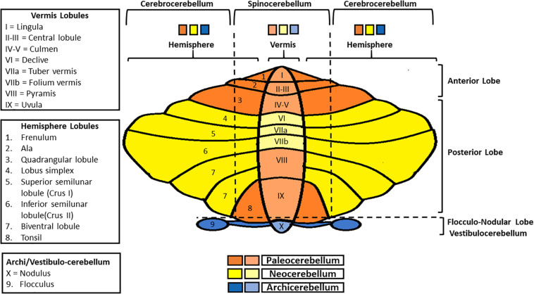
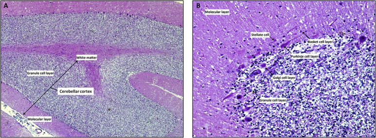
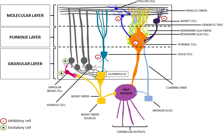
The cerebellum and its functional multiplicity and heterogeneity have been objects of curiosity and interest since ancient times, giving rise to the urge to reveal its complexity. Since the first hypothesis of cerebellar mere role in motor tuning and coordination, much more has been continuously discovered about the cerebellum's circuitry and functioning throughout centuries, leading to the currently accepted knowledge of its prominent involvement in cognitive, social, and behavioral areas. Particularly in childhood, the cerebellum may subserve several age-dependent functions, which might be compromised in several Central Nervous System pathologies. Overall, cerebellar damage may produce numerous signs and symptoms and determine a wide variety of neuropsychiatric impairments already during the evolutive age. Therefore, an early assessment in children would be desirable to address a prompt diagnosis and a proper intervention since the first months of life. Here we provide an overview of the cerebellum, retracing its morphology, histogenesis, and physiological functions, and finally outlining its involvement in typical and atypical development and the age-dependent patterns of cerebellar dysfunctions.

Keywords: age-related clinical findings; anatomy; cerebellar; cerebellum; circuitry; neurodevelopment; neuroimaging; neurophysiology.


Conflict of interest statement

The authors declare that the research was conducted in the absence of any commercial or financial relationships that could be construed as a potential conflict of interest.

Figures

FIGURE 1
FIGURE 1
Schematic representation of cerebellar gross anatomy. Anterior-posteriorly, the cerebellum presents three lobes (anterior, posterior, and flocculo-nodular), each one subdivided in lobules. Medio-laterally, it is composed of a central part (the vermis), and two lateral ones (cerebellar hemispheres). Vermis and paravermial zones form the spinocerebellum, so-called since it communicates with the spinal cord; the most lateral zones of the hemispheres constitute the cerebrocerebellum, in connection with the cerebral cortex. Finally, in regard to phylogenesis, the cerebellum can be divided into three parts: the archicerebellum (the most ancient one, corresponding to the vestibulocerebellum), paleocerebellum and neocerebellum.
FIGURE 2
FIGURE 2
The cerebellar cortex. Mid-sagittal section reveals the three layers in cerebellar folia: the superficial molecular layer, the deepest called granular and the Purkinje cells layer at the interface between the granular and molecular layers Note the inner white core of white matter (A, x4 hematoxylin/eosin stain). At higher magnification, the molecular layer contains superficially located stellate cells, basket cells which are scattered among dendritic ramifications and numerous thin axons that run parallel to the long axis of the folia. Ganglionic or Purkinje cell layer is formed of a single row of Purkinje cells with large pear-shaped bodies; while the granular layer is composed by small granule cells with dark-staining nuclei/scanty cytoplasm and Golgi type II cells (B, x20 hematoxylin/eosin stain).
FIGURE 3
FIGURE 3
Schematic representation of the cerebellar cortex. From the innermost part to the outermost one, the cerebellar cortex can be divided into three layers: the granular layer, the Purkinje layer, and the molecular layer. The former welcomes the granule cells (excitatory neurons) and the Golgi cells (inhibitory interneurons). The descending dendrites of these two cellular types together with the ascending mossy fibers (originating from the brainstem nuclei, the spinal cord and the reticular formation) make synapsis in this area, forming the so-called “glomerulus”. Purkinje cells (inhibitory neurons) are located in the middle layer, from which they send their axon to the deep nuclei, crossing the granular layer, and their dendrites to the molecular layer, forming a “dendritic tree.” Finally, basket cells and stellate cells are the two inhibitory interneurons of the molecular layer, making synapsis with the parallel fibers (originating from the T-split of the ascending axons of the granule cells).

References

    1. Abe K., Ukita H., Yorifuji S., Yanagihara T. (1997). Crossed cerebellar diaschisis in chronic Broca’s aphasia. Neuroradiology 39 624–626. 10.1007/s002340050480 - DOI - PubMed
    1. Andescavage N. N., DuPlessis A., McCarter R., Vezina G., Robertson R., Limperopoulos C. (2016). Cerebrospinal fluid and parenchymal brain development and growth in the healthy fetus. Dev. Neurosci. 38 420–429. 10.1159/000456711 - DOI - PMC - PubMed
    1. Araujo A., Carpi-Santos R., Gomes F. (2019). The role of astrocytes in the development of the cerebellum. Cerebellum 18 1017–1035. 10.1007/s12311-019-01046-0 - DOI - PubMed
    1. Ataullah A., Naqvi I. A. (2020). Cerebellar Dysfunction. London: StatPearls Publishing. - PubMed
    1. Awatramani R., Soriano P., Rodriguez C., Mai J. J., Dymecki S. M. (2003). Cryptic boundaries in roof plate and choroid plexus identified by intersectional gene activation. Nat. Genet. 35 70–75. 10.1038/ng1228 - DOI - PubMed

LinkOut - more resources