. 2021 Apr 9:12:631410.
doi: 10.3389/fimmu.2021.631410. eCollection 2021.

MR1-Restricted MAIT Cells From The Human Lung Mucosal Surface Have Distinct Phenotypic, Functional, and Transcriptomic Features That Are Preserved in HIV Infection

Affiliations

MR1-Restricted MAIT Cells From The Human Lung Mucosal Surface Have Distinct Phenotypic, Functional, and Transcriptomic Features That Are Preserved in HIV Infection

Sharon Khuzwayo et al. Front Immunol. .

Abstract

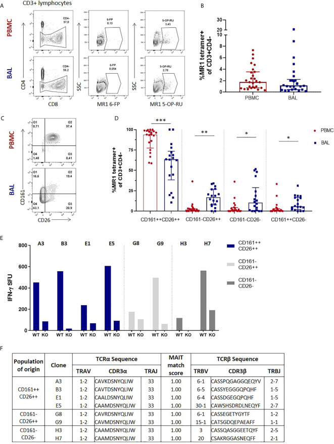
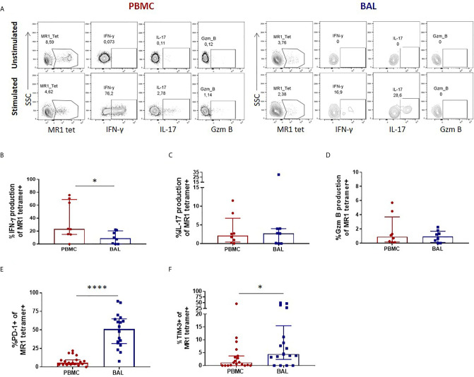
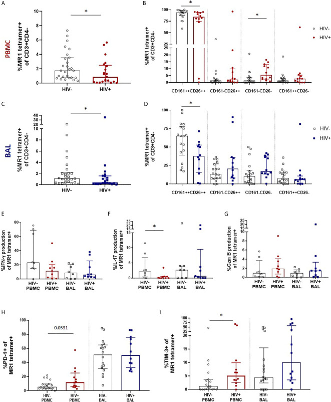
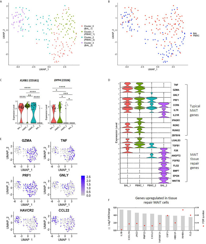
Mucosal associated invariant T (MAIT) cells are a class of innate-like T cells that utilize a semi-invariant αβ T cell receptor to recognize small molecule ligands produced by bacteria and fungi. Despite growing evidence that immune cells at mucosal surfaces are often phenotypically and functionally distinct from those in the peripheral circulation, knowledge about the characteristics of MAIT cells at the lung mucosal surface, the site of exposure to respiratory pathogens, is limited. HIV infection has been shown to have a profound effect on the number and function of MAIT cells in the peripheral blood, but its effect on lung mucosal MAIT cells is unknown. We examined the phenotypic, functional, and transcriptomic features of major histocompatibility complex (MHC) class I-related (MR1)-restricted MAIT cells from the peripheral blood and bronchoalveolar compartments of otherwise healthy individuals with latent Mycobacterium tuberculosis (Mtb) infection who were either HIV uninfected or HIV infected. Peripheral blood MAIT cells consistently co-expressed typical MAIT cell surface markers CD161 and CD26 in HIV-negative individuals, while paired bronchoalveolar MAIT cells displayed heterogenous expression of these markers. Bronchoalveolar MAIT cells produced lower levels of pro-inflammatory cytokine IFN-γ and expressed higher levels of co-inhibitory markers PD-1 and TIM-3 than peripheral MAIT cells. HIV infection resulted in decreased frequencies and pro-inflammatory function of peripheral blood MAIT cells, while in the bronchoalveolar compartment MAIT cell frequency was decreased but phenotype and function were not significantly altered. Single-cell transcriptomic analysis demonstrated greater heterogeneity among bronchoalveolar compared to peripheral blood MAIT cells and suggested a distinct subset in the bronchoalveolar compartment. The transcriptional features of this bronchoalveolar subset were associated with MAIT cell tissue repair functions. In summary, we found previously undescribed phenotypic and transcriptional heterogeneity of bronchoalveolar MAIT cells in HIV-negative people. In HIV infection, we found numeric depletion of MAIT cells in both anatomical compartments but preservation of the novel phenotypic and transcriptional features of bronchoalveolar MAIT cells.

Keywords: HIV; lung mucosal immunity; mucosal associated invariant T cells; single-cell transcriptomics; tuberculosis.


Conflict of interest statement

The authors declare that the research was conducted in the absence of any commercial or financial relationships that could be construed as a potential conflict of interest.

Figures

Figure 1
Figure 1
Phenotypic heterogeneity of bronchoalveolar MAIT cells in healthy individuals. (A) Gating strategy used to define CD3+CD4- MR1 5-OP-RU tetramer‑positive cells (using the MR1 6-FP tetramer as a control) and (B) MAIT cell frequency in the peripheral blood (PBMC) and bronchoalveolar (BAL) compartments. (C) Representative CD161 and CD26 staining of peripheral blood and bronchoalveolar CD3+CD4- MR1 tetramer‑positive MAIT cells and (D) frequency of MAIT cell phenotypic subpopulations in peripheral blood (red) and bronchoalveolar lavage (BAL) fluid (blue) (P = 0.0002, 0.0034, 0.0084 and 0.0013 respectively). MR1 tetramer‑positive T cells with atypical MAIT cell phenotype were cloned from the bronchoalveolar compartment and IFN-γ ELISpots performed to confirm MAIT cell identity. (E) MR1-dependent function of the resulting 8 bronchoalveolar MAIT cell clones (WT = M. smeg infected wild type A549 cells; KO = M. smeg infected MR1-/- A549 cells). (F) T cell receptor sequencing of bronchoalveolar MR1 tetramer-positive T cell clones grouped by phenotypic subpopulation of interest. All clones possessed the MAIT cell semi-invariant TRAV1-2/TRAJ33 alpha-chain and had MAIT match scores of 1.00 based on CDR3α sequences. Bars represent medians and error bars represent interquartile ranges. Statistical difference was determined using the Mann-Whitney U test, where *, ** and *** indicate P < 0.05, P < 0.01 and P < 0.001, respectively.
Figure 2
Figure 2
Peripheral blood MAIT cells display a pro-inflammatory profile in healthy participants. (A) Gating strategy used to define cytokine producing CD3+CD4- MR1 5-OP-RU tetramer‑positive cells observed in the unstimulated (top row) and stimulated (bottom row) conditions. Ex vivo staining of peripheral blood and bronchoalveolar MAIT cells for the production of (B) inducible IFN-γ (P = 0.0156), (C) inducible IL-17 and (D) constitutive granzyme B following 6 hour stimulation with PMA/ionomycin (n = 8). Frequency of (E) PD-1 (P < 0.0001) and (F) TIM-3 (P = 0.0417) expressing peripheral blood (red) and bronchoalveolar (blue) MAIT cells (n = 18). Bars represent medians and error bars represent interquartile ranges. Statistical difference was determined using the Mann-Whitney U test, where * and **** indicate P < 0.05 and ** P < 0.0001, respectively.
Figure 3
Figure 3
Preservation of bronchoalveolar MAIT cell phenotype and function in HIV infection. (A) MR1 tetramer staining of CD3+CD4- cells showing depletion peripheral blood MAIT cells (P = 0.0349) and (B) CD161 and CD26 staining of peripheral blood MAIT cells (P = 0.0202 and 0.0104). (C) Depletion of bronchoalveolar MAIT cells (P = 0.0471) and (D) CD161 and CD26 staining of bronchoalveolar MAIT cells (P = 0.0176) [HIV-negative (n = 18) and HIV-positive (n = 12) participants are shown]. Intracellular cytokine staining for (E) inducible IFN-γ, (F) inducible IL-17 (P = 0.0247) and (G) constitutive granzyme B production in HIV-negative (n = 8) and HIV-positive (n = 9) peripheral blood and bronchoalveolar MAIT cells following 6 hour stimulation with PMA/ionomycin. Frequency of (H) PD-1 and (I) TIM-3 (P = 0.0171) expressing MR1 tetramer-positive cells in the peripheral blood and bronchoalveolar lavage fluid of HIV-negative and HIV-positive individuals. Bars represent medians and error bars represent interquartile ranges. Statistical difference was determined using the Mann-Whitney U test, where * indicates P < 0.05.
Figure 4
Figure 4
Transcriptomic heterogeneity of MAIT cells. (A) UMAP plot showing clustering of MR1 tetramer-positive MAIT cells into four distinct transcriptomic subsets by unsupervised analysis. (B) UMAP plot showing assignment of compartment of origin to MAIT cell transcriptomic subsets. (C) Violin plot showing the expression of MAIT cell markers KLRB1 (CD161) and DPP4 (CD26) by each transcriptomic subset. (D) Violin plot showing the expression of typical MAIT cell genes (top), and tissue repair genes (bottom) by MR1 tetramer-positive MAIT cells from each transcriptomic subset. (E) UMAP plots showing genes of interest expressed by BAL_2 MAIT cells, including MAIT effector genes and tissue repair genes. (F) Bulk RNA-sequence analysis showing the upregulation of MAIT tissue repair genes in the bronchoalveolar MAIT cells of HIV-negative individuals as compared to peripheral blood MAIT cells, where *, **, *** and **** indicate P < 0.05, P < 0.01, P < 0.001 and P < 0.0001, respectively.

Similar articles

Cited by

References

    1. Tilloy F, Treiner E, Park S-H, Garcia C, Lemonnier F, de la Salle H, et al. . An invariant T cell receptor α chain defines a novel TAP-independent major histocompatibility complex class Ib–restricted α/β T cell subpopulation in mammals. J Exp Med (1999) 189(12):1907–21. 10.1084/jem.189.12.1907 - DOI - PMC - PubMed
    1. Treiner E, Duban L, Bahram S, Radosavljevic M, Wanner V, Tilloy F, et al. . Selection of evolutionarily conserved mucosal-associated invariant T cells by MR1. Nature (2003) 422(6928):164–9. 10.1038/nature01433 - DOI - PubMed
    1. Porcelli S, Yockey CE, Brenner MB, Balk SP. Analysis of T cell antigen receptor (TCR) expression by human peripheral blood CD4-8- alpha/beta T cells demonstrates preferential use of several V beta genes and an invariant TCR alpha chain. J Exp Med (1993) 178(1):1–16. 10.1084/jem.178.1.1 - DOI - PMC - PubMed
    1. Gold MC, Cerri S, Smyk-Pearson S, Cansler ME, Vogt TM, Delepine J, et al. . Human mucosal associated invariant T cells detect bacterially infected cells. PloS Biol (2010) 8(6):e1000407. 10.1371/journal.pbio.1000407 - DOI - PMC - PubMed
    1. Kjer-Nielsen L, Patel O, Corbett AJ, Le Nours J, Meehan B, Liu L, et al. . MR1 presents microbial vitamin B metabolites to MAIT cells. Nature (2012) 491(7426):717–23. 10.1038/nature11605 - DOI - PubMed

Publication types

MeSH terms

Substances