

Macrophage-Mediated Tissue Vascularization: Similarities and Differences Between Cornea and Skin
- PMID: 33897716
- PMCID: PMC8058454
- DOI: 10.3389/fimmu.2021.667830
Macrophage-Mediated Tissue Vascularization: Similarities and Differences Between Cornea and Skin
Abstract
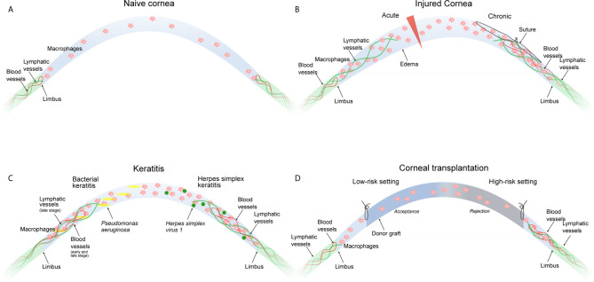
Macrophages are critical mediators of tissue vascularization both in health and disease. In multiple tissues, macrophages have been identified as important regulators of both blood and lymphatic vessel growth, specifically following tissue injury and in pathological inflammatory responses. In development, macrophages have also been implicated in limiting vascular growth. Hence, macrophages provide an important therapeutic target to modulate tissue vascularization in the clinic. However, the molecular mechanisms how macrophages mediate tissue vascularization are still not entirely resolved. Furthermore, mechanisms might also vary among different tissues. Here we review the role of macrophages in tissue vascularization with a focus on their role in blood and lymphatic vessel formation in the barrier tissues cornea and skin. Comparing mechanisms of macrophage-mediated hem- and lymphangiogenesis in the angiogenically privileged cornea and the physiologically vascularized skin provides an opportunity to highlight similarities but also tissue-specific differences, and to understand how macrophage-mediated hem- and lymphangiogenesis can be exploited for the treatment of disease, including corneal wound healing after injury, graft rejection after corneal transplantation or pathological vascularization of the skin.
Keywords: angiogenesis; cornea; lymphangiogenesis; macrophages; monocytes; skin.
Copyright © 2021 Hadrian, Willenborg, Bock, Cursiefen, Eming and Hos.
Conflict of interest statement
The authors declare that the research was conducted in the absence of any commercial or financial relationships that could be construed as a potential conflict of interest.
Figures
Similar articles
-
VEGF-A expression by HSV-1-infected cells drives corneal lymphangiogenesis.J Exp Med. 2010 Jan 18;207(1):101-15. doi: 10.1084/jem.20091385. Epub 2009 Dec 21. J Exp Med. 2010. PMID: 20026662 Free PMC article.
-
The maintenance of lymphatic vessels in the cornea is dependent on the presence of macrophages.Invest Ophthalmol Vis Sci. 2012 May 31;53(6):3145-53. doi: 10.1167/iovs.11-8010. Invest Ophthalmol Vis Sci. 2012. PMID: 22511631
-
Time course of angiogenesis and lymphangiogenesis after brief corneal inflammation.Cornea. 2006 May;25(4):443-7. doi: 10.1097/01.ico.0000183485.85636.ff. Cornea. 2006. PMID: 16670483
-
Corneal lymphangiogenesis: implications in immunity.Semin Ophthalmol. 2009 May-Jun;24(3):135-8. doi: 10.1080/08820530902801320. Semin Ophthalmol. 2009. PMID: 19437348 Review.
-
Lymphangiogenesis Guidance Mechanisms and Therapeutic Implications in Pathological States of the Cornea.Cells. 2023 Jan 14;12(2):319. doi: 10.3390/cells12020319. Cells. 2023. PMID: 36672254 Free PMC article. Review.
Cited by
-
Translational frontiers: insight from lymphatics in skin regeneration.Front Physiol. 2024 Feb 29;15:1347558. doi: 10.3389/fphys.2024.1347558. eCollection 2024. Front Physiol. 2024. PMID: 38487264 Free PMC article. Review.
-
Signal regulatory protein α dynamically mediates macrophage polarization facilitated alleviation of ischemic diseases.Cell Biosci. 2024 Dec 20;14(1):150. doi: 10.1186/s13578-024-01325-2. Cell Biosci. 2024. PMID: 39707436 Free PMC article.
-
Ontogenesis of the Mouse Ocular Surface Lymphatic Vascular Network.Invest Ophthalmol Vis Sci. 2023 Dec 1;64(15):7. doi: 10.1167/iovs.64.15.7. Invest Ophthalmol Vis Sci. 2023. PMID: 38054922 Free PMC article.
-
Sulfur mustard corneal injury is associated with alterations in the epithelial basement membrane and stromal extracellular matrix.Exp Mol Pathol. 2022 Oct;128:104807. doi: 10.1016/j.yexmp.2022.104807. Epub 2022 Jul 4. Exp Mol Pathol. 2022. PMID: 35798063 Free PMC article.
-
The Role of Connexin in Ophthalmic Neovascularization and the Interaction between Connexin and Proangiogenic Factors.J Ophthalmol. 2022 Jun 22;2022:8105229. doi: 10.1155/2022/8105229. eCollection 2022. J Ophthalmol. 2022. PMID: 35783340 Free PMC article. Review.
References
Publication types
MeSH terms
LinkOut - more resources
Full Text Sources
Other Literature Sources