

Clival prolactinoma masquerading as a chordoma: a case report
- PMID: 33897924
- PMCID: PMC8053778
- DOI: 10.1016/j.radcr.2021.02.071
Clival prolactinoma masquerading as a chordoma: a case report
Abstract
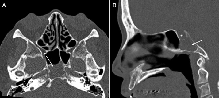
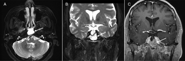
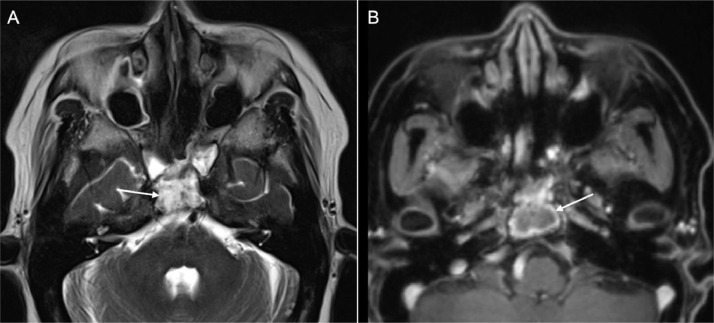
Ectopic pituitary adenomas are a rare clinical entity and are frequently mistaken for other base of skull lesions on imaging. We report the clinical presentation and management of a woman presenting with an ectopic prolactinoma located in the clivus. A 66-year-old female presented with a 6-month history of headaches and light-headedness. Anatomical imaging demonstrated a clival lesion most suspicious for chordoma. Endocrinological assessment revealed modestly increased prolactin level with lower-than-expected gonadotrophins levels for her age. Surgical resection confirmed an ectopic prolactinoma. A skull base lesion in a patient with hormonal derangement should lend to a high clinical suspicion of an EPA as they may be treated with medications before surgery. Guidelines could assist clinicians investigating skull-based lesions to identify the rare, but important diagnosis of ectopic pituitary adenomas.
Keywords: Adenoma or tumor; Chordoma; Clival; Ectopic; Pituitary.
Crown Copyright © 2021 Published by Elsevier Inc. on behalf of University of Washington.
Figures
Similar articles
-
Clival Ectopic Pituitary Adenoma Mimicking a Chordoma: Case Report and Review of the Literature.Case Rep Neurol Med. 2016;2016:8371697. doi: 10.1155/2016/8371697. Epub 2016 Jan 13. Case Rep Neurol Med. 2016. PMID: 26885420 Free PMC article.
-
Ectopic dopamine agonist-resistant macroprolactinoma to the clivus masquerading as a chordoma - A case report.Endocr Regul. 2025 Mar 12;59(1):10-16. doi: 10.2478/enr-2025-0002. Print 2025 Jan 1. Endocr Regul. 2025. PMID: 40073404
-
Ectopic pituitary adenomas: common presentations of a rare entity.Pituitary. 2019 Aug;22(4):339-343. doi: 10.1007/s11102-019-00954-y. Pituitary. 2019. PMID: 30895500
-
Ectopic invasive ACTH-secreting pituitary adenoma mimicking chordoma: a case report and literature review.BMC Neurol. 2023 Feb 23;23(1):81. doi: 10.1186/s12883-023-03124-7. BMC Neurol. 2023. PMID: 36814261 Free PMC article. Review.
-
[Unilateral exophthalmos caused by a prolactin producing ectopic pituitary adenoma: case report].No Shinkei Geka. 2002 Jun;30(6):623-8. No Shinkei Geka. 2002. PMID: 12094689 Review. Japanese.
Cited by
-
Hyperprolactinemia caused by extra-pituitary prolactin secretion: a systematic review.Endocr Connect. 2025 Jul 1;14(7):e250054. doi: 10.1530/EC-25-0054. Print 2025 Jul 1. Endocr Connect. 2025. PMID: 40531582 Free PMC article.
-
Clinical and radiological presentation of parasellar ectopic pituitary adenomas: case series and systematic review of the literature.J Endocrinol Invest. 2022 Aug;45(8):1465-1481. doi: 10.1007/s40618-022-01758-x. Epub 2022 Feb 11. J Endocrinol Invest. 2022. PMID: 35147925
References
Publication types
LinkOut - more resources
Full Text Sources
Other Literature Sources
Research Materials
