Case Reports
. 2021 Apr 9;16(6):1363-1367.
doi: 10.1016/j.radcr.2021.03.045. eCollection 2021 Jun.

A rare case of lipomatous pseudohypertrophy of the pancreas

Affiliations
Case Reports

A rare case of lipomatous pseudohypertrophy of the pancreas

Vu Dang Luu et al. Radiol Case Rep. .

Abstract

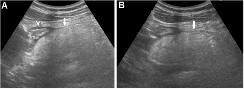
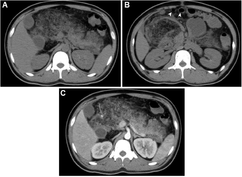
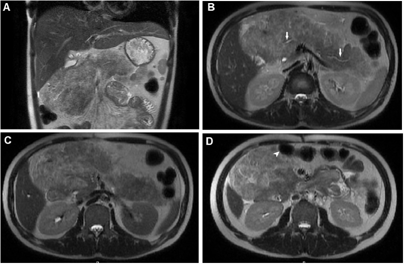
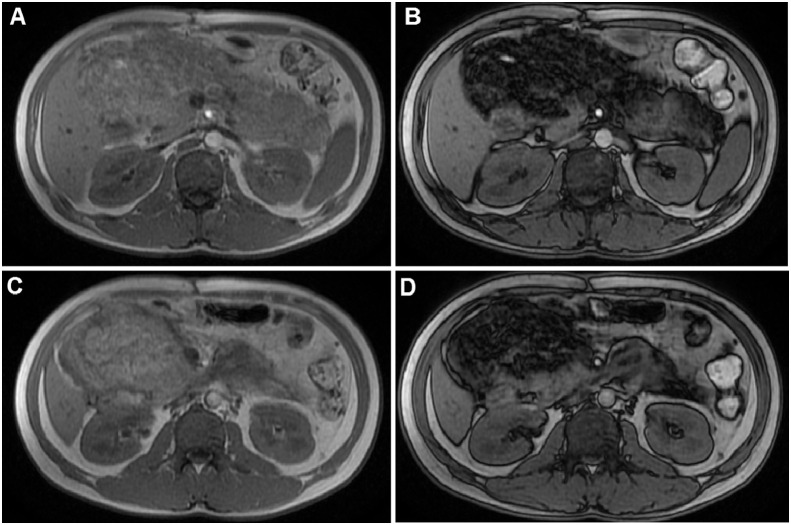
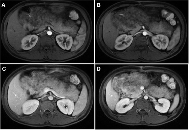
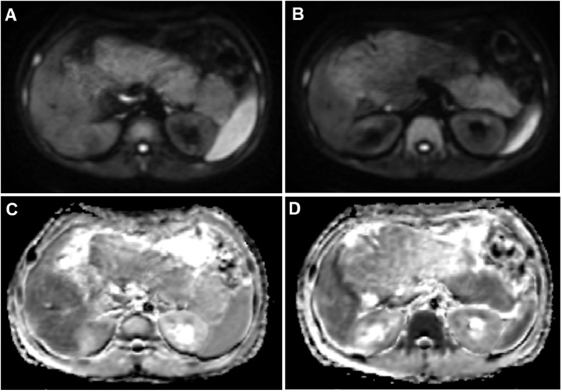
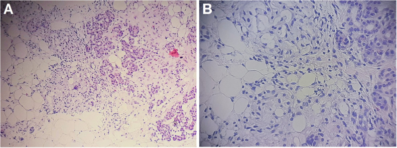
Lipomatous pseudohypertrophy of the pancreas is a rare disease with unknown etiology, and the pancreas parenchyma is replaced by pancreatic parenchyma by fat tissue. In this article, we aimed to report the case of a 26-year-old male patient admitted to hospital with loss of appetite for 6 months. Abdominal computed tomography (CT) and magnetic resonance imaging (MRI) scans showed diffuse enlargement and fatty replacement over the whole pancreas, with scattered remnants of pancreatic parenchyma. Histologic results defined lipomatous pseudohypertrophy of the pancreas. To summarize, this case report is to put forward this extremely rare presentation and to sensitize clinicians that this entity can be a cause of exocrine pancreatic insufficiency, which requires patient follow-up for the appropriate treatment.

Keywords: Lipomatous pseudohypertrophy; Pancreas; Pancreas lipomatosis.


Figures

Fig 1
Fig. 1
Abdominal ultrasound showing that the normal pancreas was replaced with a hyperechoic and well-defined border mass (A and B) (arrow), which compressed the duodenum (A) (arrowhead).
Fig 2
Fig. 2
Abdominal CT scans. (A and B) CT scans, pre-contrast, revealed that the pancreas was enlarged, well-circumscribed, and replaced with low-density fat tissue. (C) CT scans, post-contrast, showed that the normal parenchyma enhanced. The largest diameter was observed in the head, at 135 mm, and the length of the pancreas was 220 mm. The small bowel had been displaced anteriorly (arrowhead).
Fig 3
Fig. 3
T2-weighted MRI. T2-weighted coronal (A) and T2-weighted axial (B, C and D) images showed that the pancreas was enlarged and well-circumscribed and the parenchymal signal was heterogeneous, with several hyperintense areas. The main pancreatic duct was normal (B) (arrow). The pancreas displaced the small bowel anteriorly (D) (arrowhead).
Fig 4
Fig. 4
T1-weighted MRI. (A and C) In-phase T1-weighted images showed that the pancreas was enlarged with heterogeneous, with high signal intensity. (B and D) Opposed-phase T1-weighted images showed that parts of the parenchyma lost signal intensity due to adipose tissue replacement.
Fig 5
Fig. 5
T1-weighted MRI with contrast on arterial (A and B) and venous phases (C and D) displayed the enhancement of preserved normal parenchyma; fat tissue was unenhanced.
Fig 6
Fig. 6
Diffusion-weighted imaging (A and B) and ADC map imaging (C and D) showed that the pancreatic parenchyma was hypointense on diffusion-weighted images and had similar ADC values as the spleen.
Fig 7
Fig. 7
Microscopic images of the pancreas, visualized with hematoxylin and eosin (HE) stain. (A) HE staining, ×100. (B) HE staining, ×200. The pathological examination revealed the diffuse replacement of the normal pancreatic parenchyma with mature adipose tissue. Scattered, residual pockets of normal pancreatic acinar cells were observed. The pancreatic ductal system remained intact.

References

    1. Hantelmann W. Fettsucht und Atrophie der Bauchspecicheldruse bei Jungendlichen. Virchows Arch. 1931;282:630–642.
    1. Siegler D.I. Lipomatous pseudohypertrophy of the pancreas associated with chronic pulmonary suppuration in an adult. Postgrad Med J. 1974;50(579):53–55. - PMC - PubMed
    1. Altinel D., Basturk O., Sarmiento J.M., Martin D., Jacobs M.J., Kooby D.A. Lipomatous pseudohypertrophy of the pancreas: a clinicopathologically distinct entity. Pancreas. 2010;39:392–397. - PMC - PubMed
    1. Yasuda M., Niina Y., Uchida M., Fujimori N., Nakamura T., Oono T. A case of lipomatous pseudohypertrophy of the pancreas diagnosed by typical imaging. Jop. 2010;11(4):385–388. - PubMed
    1. Shimada M., Shibahara K., Kitamura H., Demura Y., Hada M., Takehara A. Lipomatous pseudohypertrophy of the pancreas taking the form of huge massive lesion of the pancreatic head. Case Rep Gastroenterol. 2010;4(3):457–464. - PMC - PubMed

Publication types

LinkOut - more resources