. 2021 Apr 9:2021:6673610.
doi: 10.1155/2021/6673610. eCollection 2021.

Electroacupuncture Alleviates Inflammation of Dry Eye Diseases by Regulating the α 7nAChR/NF- κ B Signaling Pathway

Affiliations

Electroacupuncture Alleviates Inflammation of Dry Eye Diseases by Regulating the α 7nAChR/NF- κ B Signaling Pathway

Ning Ding et al. Oxid Med Cell Longev. .

Abstract

Purpose: We tried to investigate whether electroacupuncture (EA) can reduce inflammation of dry eye disease (DED) by regulating α7nAChR and inhibiting the NF-κB signaling pathway.

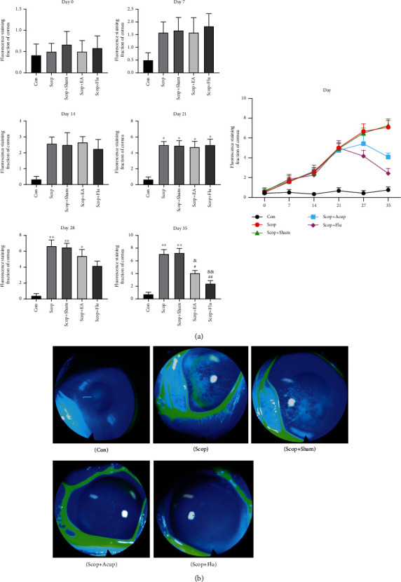
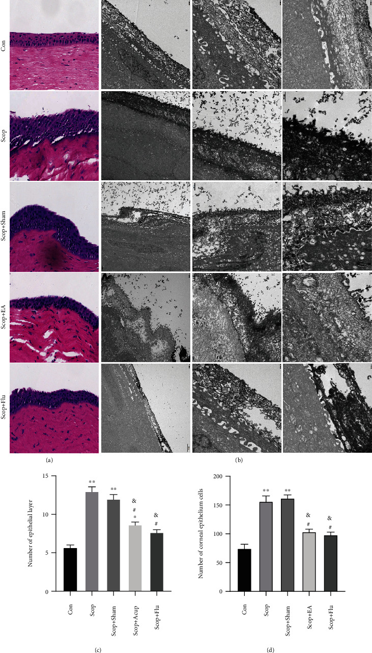
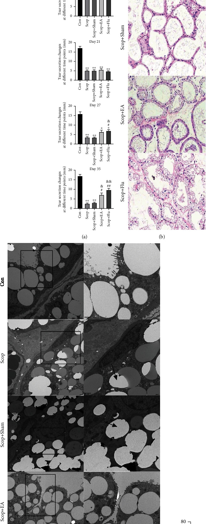
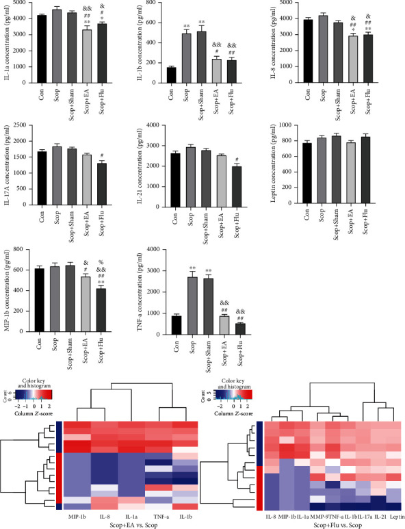
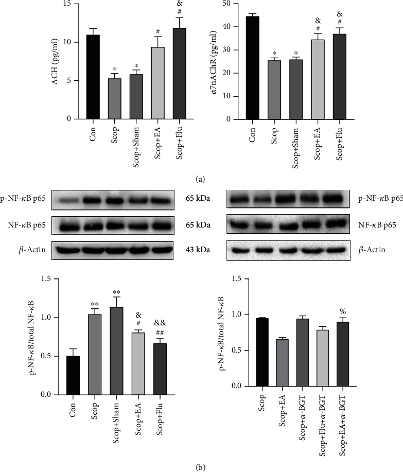
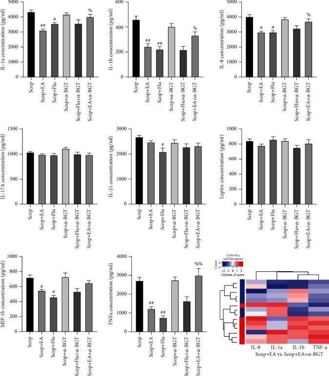
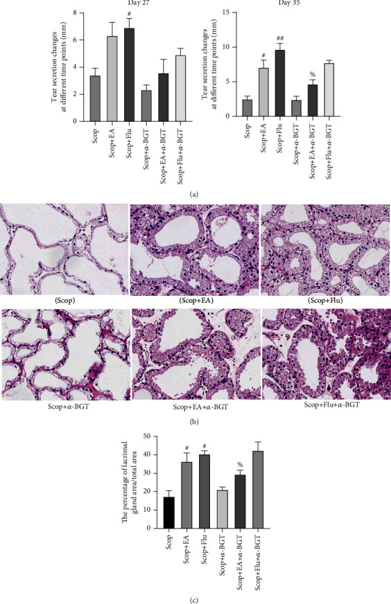
Methods: Healthy New Zealand white rabbits were treated with scopolamine hydrobromide (Scop) for 21 consecutive days to establish the DED animal model. After 21 days, EA, fluorometholone (Flu), and α7nAChR antagonist (α-BGT) treatments were performed, and the Scop injection was continued until day 35. During treatment, the fluorescence staining of the corneal epithelium and the level of tear flow were observed. The influence of EA on the LG pathology and inflammatory factors ACh, α7nAChR, and NF-κB was detected using the LG histopathology, transmission electron microscopy (TEM), cytokine protein chip technology, enzyme-linked immunosorbent assay (ELISA), and Western blot.

Results: The EA stimulation can reduce the corneal epithelial damage and repair epithelial cell ultrastructure, promote the tear secretion, relieve the LG atrophy and decrease lipid droplet accumulation in LG acinar cell, and reduce the levels of inflammatory cytokines (i.e., IL-1, MIP-1b, TNF-α, and IL-8) in the LG. The protective effect of EA on the inflammation and the ocular surface is similar to the corticosteroid Flu. EA and Flu can upregulate the expression of the α7nAChR and downregulate the expression of NF-κB. The α7nAChR antagonist α-BGT can reverse the protective effect of EA on the LG and the inhibitory effect on the NF-κB pathway and the expression of inflammatory factors but cannot affect the expression of Flu on the NF-κB pathway and inflammatory factors.

Conclusion: These results prove that EA can alleviate DEDs by stimulating the acupoints around the eyes. These beneficial effects are related to the upregulation of α7nAChR and the downregulation of NF-κB in the LG. The protective effect of LG is mediated through the anti-inflammatory pathway mediated by α7nAChR. EA can reduce the NF-κB P65 nuclear transcription and reduce inflammatory factors by regulating α7nAChR. This expression indicates that the α7nAChR/NF-κB signaling pathway may serve as a potential therapeutic target for EA to treat DEDs.


Conflict of interest statement

The authors declare no conflict of interest.

Figures

Figure 1
Figure 1
Effect of the electroacupuncture treatment on the corneal fluorescence staining in DED induced by scopolamine hydrobromide: (a) corneal fluorescence staining score; (b) corneal fluorescence staining on day 35. Quantitative data are expressed as mean ± SEM (n = 6). The Scop group has increased corneal fluorescence staining score significantly on day 21 when compared with the Con group. After 14 days of treatment, the EA and Scop+Flu groups have decreased corneal fluorescence staining score when compared with the Scop group, indicating that the corneal epithelial damage has been improved. P < 0.05 and ∗∗P < 0.01 vs. the Con group; #P < 0.05 and ##P < 0.01 vs. the Scop group; &&P < 0.01 and &&P < 0.01 vs. the Scop+Sham group.
Figure 2
Figure 2
Alterations to the corneal epithelium following treatment. (a) Representative images demonstrating epithelial layers and epithelium cells in the cornea (hematoxylin-eosin staining, 20). (b) Corneal epithelial cells under transmission electron microscopy. (c) The number of epithelial layers. (d) The number of cells in the cornea epithelium was determined. P < 0.05 and ∗∗P < 0.01 vs. the Con group; #P < 0.05 vs. the Scop group; &P < 0.05 vs. the Scop+Sham group.
Figure 3
Figure 3
Effect of the electroacupuncture treatment on the tear flow and the lacrimal glands in the DED induced by scopolamine hydrobromide: (a) tear fluid flow (n = 6); (b) histopathological images of the cornea (hematoxylin-eosin staining, ×20) on day 35 (n = 3); (c) lacrimal glands under transmission electron microscopy; (d) the percentage of lacrimal gland area/total area (%). Quantitative data are expressed as mean ± SEM. P < 0.05, ∗∗P < 0.01, and ∗∗∗P < 0.001 vs. the Con group; #P < 0.05 and ##P < 0.01 vs. the Scop group; &P < 0.05 and &&P < 0.01 vs. the Scop+Sham group. (c) Ultrastructural analysis under transmission electron microscopy revealed that EA decreased lipid droplet accumulation in LG acinar cell. Lipid droplets (black arrows).
Figure 4
Figure 4
Changes in the cytokines and the chemokines in rabbit LGs on day 35. Quantitative data are expressed as mean ± SEM (n = 4). EA stimulus significantly decreased the levels of IL-1a, IL-1b, MIP-1b, TNF-α, and IL-8 when compared with the Scop group. The Scop+Flu group significantly decreased the levels of IL-1a, IL-1b, IL-17A, IL-21, MIP-1b, TNF-α, and IL-8 when compared with the Scop group. P < 0.05 and ∗∗P < 0.01 vs. the Con group; #P < 0.05 and ##P < 0.01 vs. the Scop group; &P < 0.05 and &&P < 0.01 vs. the Scop+Sham group; %P < 0.05 vs. the Scop+EA group. Clustering heat map: blue represents the Scop group.
Figure 5
Figure 5
Effect of electroacupuncture on the changes in ACH and α7nAChR in the LG determined using ELISA. (a) Western blot of the effect of EA on the activation of NF-κB with β-actin as a load control. (b) Quantitative data are expressed as mean ± SEM (n = 3). P < 0.05 and ∗∗P < 0.01 vs. the Con group; #P < 0.05 and ##P < 0.01 vs. the Scop group; &P < 0.05 and &&P < 0.01 vs. the Scop+Sham group; %P < 0.05 vs. the Scop+EA group.
Figure 6
Figure 6
Dependence of the regulation of the electroacupuncture stimulation on inflammatory factors and chemokines on α7nAChR. The α7nAChR antagonist reverses the inhibitory effect of the EA stimulation on proinflammatory cytokines and chemokines. Quantitative data are expressed as mean ± SEM (n = 4). #P < 0.05 and ##P < 0.01 vs. the Scop group; %P < 0.05 and %%P < 0.01 vs. the Scop+EA group. Clustering heat map: blue represents the EA group.
Figure 7
Figure 7
α7nAChR is involved in the protective effect of EA on the LG: (a) tear fluid flow (n = 6); (b) histopathological images of the cornea (hematoxylin-eosin staining, ×20) on day 35 (n = 3); (c) the percentage of lacrimal gland area/total area (%). Quantitative data are expressed as mean ± SEM. #P < 0.05 and ##P < 0.01 vs. the Scop group; %P < 0.05 vs. the Scop+EA group.

References

    1. Jones L., Downie L. E., Korb D., et al. TFOS DEWS II management and therapy report. The Ocular Surface. 2017;15(3):575–628. doi: 10.1016/j.jtos.2017.05.006. - DOI - PubMed
    1. Stapleton F., Alves M., Bunya V. Y., et al. TFOS DEWS II epidemiology report. The Ocular Surface. 2017;15(3):334–365. doi: 10.1016/j.jtos.2017.05.003. - DOI - PubMed
    1. Peri Y., Agmon-Levin N., Theodor E., Shoenfeld Y. Sjogren’s syndrome, the old and the new. Best Practice & Research: Clinical Rheumatology. 2012;26(1):105–117. doi: 10.1016/j.berh.2012.01.012. - DOI - PubMed
    1. Zoukhri D. Effect of inflammation on lacrimal gland function. Experimental Eye Research. 2006;82(5):885–898. doi: 10.1016/j.exer.2005.10.018. - DOI - PMC - PubMed
    1. Dursun D., Wang M., Monroy D., et al. A mouse model of keratoconjunctivitis sicca. Investigative Ophthalmology & Visual Science. 2002;43(3):632–638. - PubMed

LinkOut - more resources