. 2020 Nov 20;8(2):209-221.
doi: 10.1093/nop/npaa075. eCollection 2021 Apr.

FGFR3 overexpression is a useful detection tool for FGFR3 fusions and sequence variations in glioma

Affiliations

FGFR3 overexpression is a useful detection tool for FGFR3 fusions and sequence variations in glioma

Jens Schittenhelm et al. Neurooncol Pract. .

Abstract

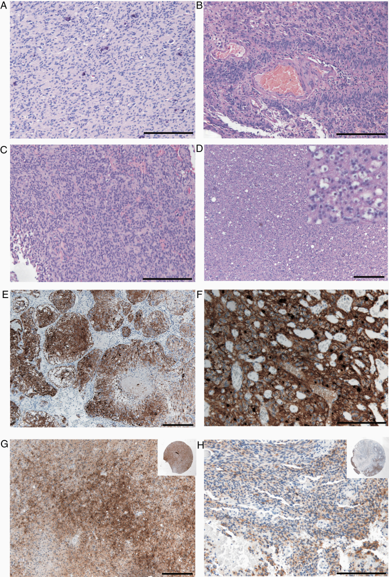
Background: Fibroblast growth factor receptor (FGFR) inhibitors are currently used in clinical development. A subset of glioblastomas carries gene fusion of FGFR3 and transforming acidic coiled-coil protein 3. The prevalence of other FGFR3 alterations in glioma is currently unclear.

Methods: We performed RT-PCR in 101 glioblastoma samples to detect FGFR3-TACC3 fusions ("RT-PCR cohort") and correlated results with FGFR3 immunohistochemistry (IHC). Further, we applied FGFR3 IHC in 552 tissue microarray glioma samples ("TMA cohort") and validated these results in two external cohorts with 319 patients. Gene panel sequencing was carried out in 88 samples ("NGS cohort") to identify other possible FGFR3 alterations. Molecular modeling was performed on newly detected mutations.

Results: In the "RT-PCR cohort," we identified FGFR3-TACC3 fusions in 2/101 glioblastomas. Positive IHC staining was observed in 73/1024 tumor samples of which 10 were strongly positive. In the "NGS cohort," we identified FGFR3 fusions in 9/88 cases, FGFR3 amplification in 2/88 cases, and FGFR3 gene mutations in 7/88 cases in targeted sequencing. All FGFR3 fusions and amplifications and a novel FGFR3 K649R missense mutation were associated with FGFR3 overexpression (sensitivity and specificity of 93% and 95%, respectively, at cutoff IHC score > 7). Modeling of these data indicated that Tyr647, a residue phosphorylated as a part of FGFR3 activation, is affected by the K649R mutation.

Conclusions: FGFR3 IHC is a useful screening tool for the detection of FGFR3 alterations and could be included in the workflow for isocitrate dehydrogenase (IDH) wild-type glioma diagnostics. Samples with positive FGFR3 staining could then be selected for NGS-based diagnostic tools.

Keywords: FGFR3; glioma; panel sequencing; targeted treatment.


Figures

Figure 1.
Figure 1.
CONSORT diagram depicting samples used in the study. A immunoreactive (IR) score > 7 corresponds to overexpression in FGFR3 immunohistochemistry evaluation results. Abbreviations: NGS = next-generation sequencing; TMA = tissue microarray; RT-PCR = reverse transcriptase PCR; IHC = immunohistochemistry; FGFR3 = fibroblast growth factor receptor 3.
Figure 2.
Figure 2.
FGFR3 fusions in glioblastoma: A, RT-PCR product for FGFR3 exon 17-TACC3 exon 10 primer pairs. B, Confirmatory Sanger sequence of FGFR3-TACC breakpoint in Case #1. C, FGFR3 (left column) and TACC3 (right column) immunohistochemistry in a FGFR-TACC3 fusion-positive (Case #1) and fusion-negative (Case #4) tumor. D, Full mounted slide staining confirming strong FGFR3 immunoreactivity score (IRS: 12, insets: full photo of stained TMA punch, scale bars: 200 µm).
Figure 3.
Figure 3.
FGFR3 immunohistochemistry distribution among the glioma tumor cohorts analyzed in this study. Positive FGFR3 staining was observed in 73 out of 1024 samples total. FGFR3 overexpression (scores > 7, Figure 1) was detected in 26 samples. Strong FGFR3 staining was observed in 1% of all tumors analyzed (10/1024) and was significantly associated with the presence of a FGFR3-TACC3 fusion.
Figure 4.
Figure 4.
Summary of genetic profiles found in FGFR3-altered tumors (yellow box: protein-affecting mutation, violet amp: amplification, brown del: deletion, MGMT promotor M: methylated, U: unmethylated, blue loss: nuclear expression absent, *: IHC data from primary tumor). The FGFR3-TACC3 fusions in Cases #1 and #3 were also identified by RT-PCR.
Figure 5.
Figure 5.
Histological patterns observed in FGFR3-positive tumors as reported by Bielle et al.A, Monomorphous ovoid nuclei with focal microcalcifications (HE). B, Attachment of tumor cells to vessels by thin cytoplasmic processes resembling vague pseudorosettes. C, Organization of tumor cells in pseudo-ependymal pseudorosettes (HE). D, Isolated tumor cells surrounded by pale ovoid cytoplasm (HE, inset higher magnification). E, Organoid nesting of tumor cells (FGFR3 immunohistochemistry) with surrounding vascularization. F, Endocrinoid network of thin capillaries and tumor cells with pale cytoplasm and membranous enrichment of FGFR3 immunohistochemistry. G, Primary tumor with moderate FGFR3 immunohistochemistry (IRS score 6) and reduced expression in tumor recurrence one year later (IRS score 2, insets show corresponding stained tissue microarray punches). Scale bars: 200 µm.

References

    1. Claes A, Idema AJ, Wesseling P. Diffuse glioma growth: a guerilla war. Acta Neuropathol. 2007;114(5):443–458. - PMC - PubMed
    1. Watanabe T, Nobusawa S, Kleihues P, et al. IDH1 mutations are early events in the development of astrocytomas and oligodendrogliomas. Am J Pathol. 2009;174(4):1149–1153. - PMC - PubMed
    1. Brennan CW, Verhaak RG, McKenna A, et al. The somatic genomic landscape of glioblastoma. Cell. 2013;155(2):462–477. - PMC - PubMed
    1. Phillips HS, Kharbanda S, Chen R, et al. Molecular subclasses of high-grade glioma predict prognosis, delineate a pattern of disease progression, and resemble stages in neurogenesis. Cancer Cell. 2006;9(3):157–173. - PubMed
    1. Capper D, Jones DTW, Sill M, et al. DNA methylation-based classification of central nervous system tumours. Nature. 2018;555(7697):469–474. - PMC - PubMed