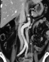
Inferior Vena Cava Filter Placement in a Duplicated Inferior Vena Cava: A Case Report
- PMID: 33898058
- PMCID: PMC8053213
- DOI: 10.5001/omj.2021.29
Inferior Vena Cava Filter Placement in a Duplicated Inferior Vena Cava: A Case Report
Abstract
Inferior vena cava (IVC) duplication is a well-known anatomic variation that is important when relevant procedures are being planned. Duplication of IVC is a relatively rare to detect especially vascular anomaly with a prevalence of 1.5% (range 0.2-3.0%). Knowing this anatomical variation is very important in cases of IVC filter placement. Filter placement in duplicated IVC cases has many options like placing it in both vena cavae, suprarenal filter placement, or coil embolization of the intervenous segment plus placing a filter in the right IVC. We report a case of a patient with newly diagnosed bladder cancer who had a high risk of thrombosis and a recent massive pulmonary embolism. The patient was planned for transurethral resection of the bladder tumor. As a prophylactic measure, an IVC filter placement was requested to prevent further pulmonary emboli that might occur during or after surgery. Cavography showed a duplicated IVC, and the filter placement was performed in the suprarenal portion and was proved to be an adequate and safe procedure. No procedure-related complications were reported. There are few worldwide reported cases of filter placement in a duplicated vena cava, and to our best knowledge, this is the first case reported in Oman.
Keywords: Anatomic Variation; Oman; Renal Veins; Vena Cava, Inferior.
The OMJ is Published Bimonthly and Copyrighted 2021 by the OMSB.
Figures


References
-
- Malgor RD, Sobreira ML, Boaventura PN, Moura R, Yoshida WB. Filter placement in duplicated inferior vena cava: case report and review of the literature. J Vasc Bras 2008;7(2):167-170 . 10.1590/S1677-54492008000200013 - DOI
LinkOut - more resources
Full Text Sources
Other Literature Sources
Research Materials
