. 2021 Apr 9:11:639980.
doi: 10.3389/fonc.2021.639980. eCollection 2021.

LncRNA LIPE-AS1 Predicts Poor Survival of Cervical Cancer and Promotes Its Proliferation and Migration via Modulating miR-195-5p/MAPK Pathway

Affiliations

LncRNA LIPE-AS1 Predicts Poor Survival of Cervical Cancer and Promotes Its Proliferation and Migration via Modulating miR-195-5p/MAPK Pathway

Jie Zhang et al. Front Oncol. .

Retraction in

Abstract

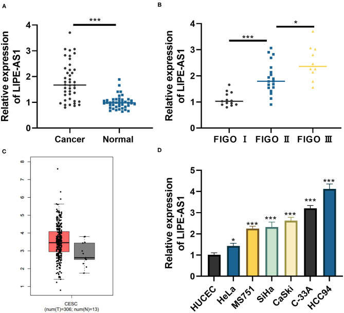
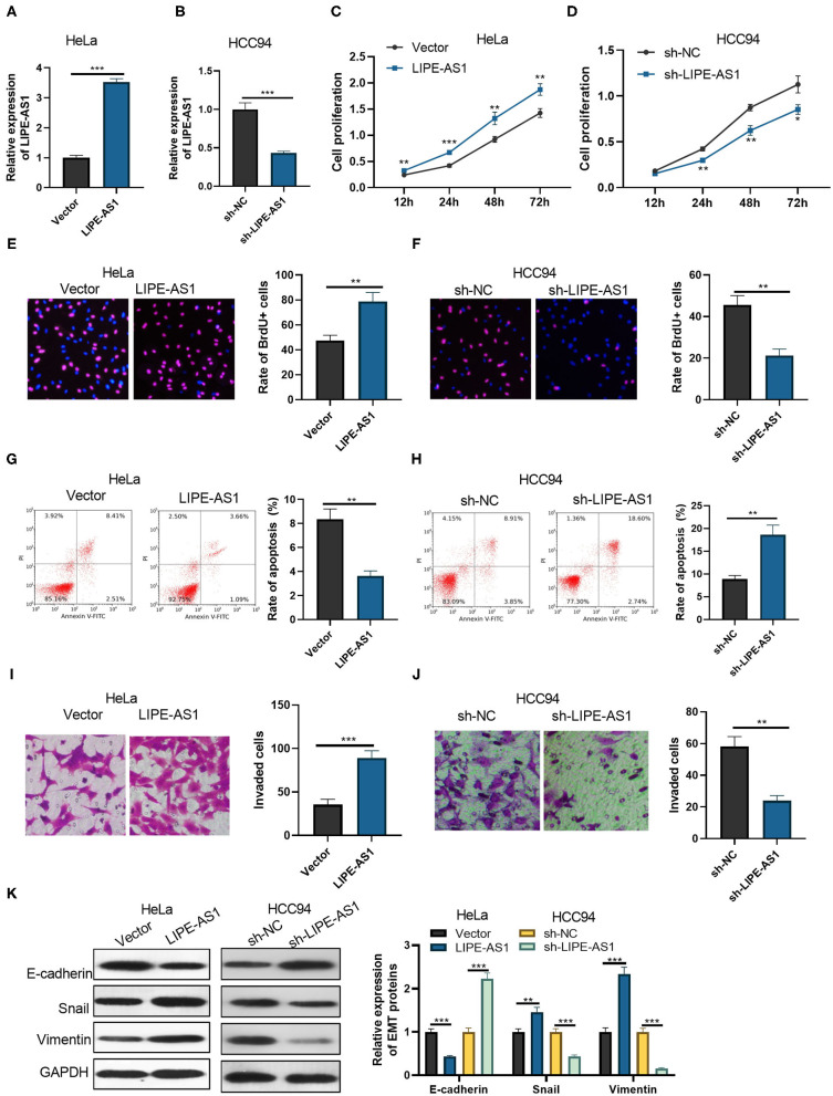
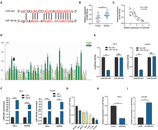
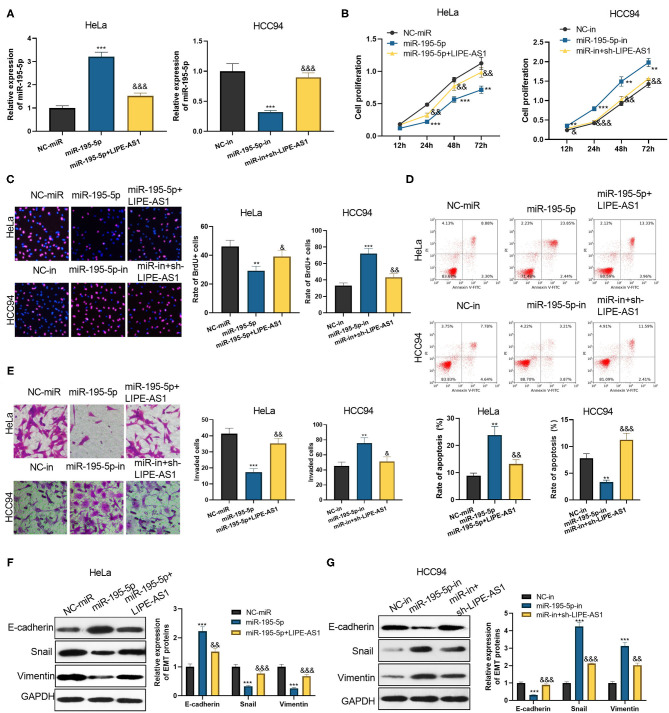
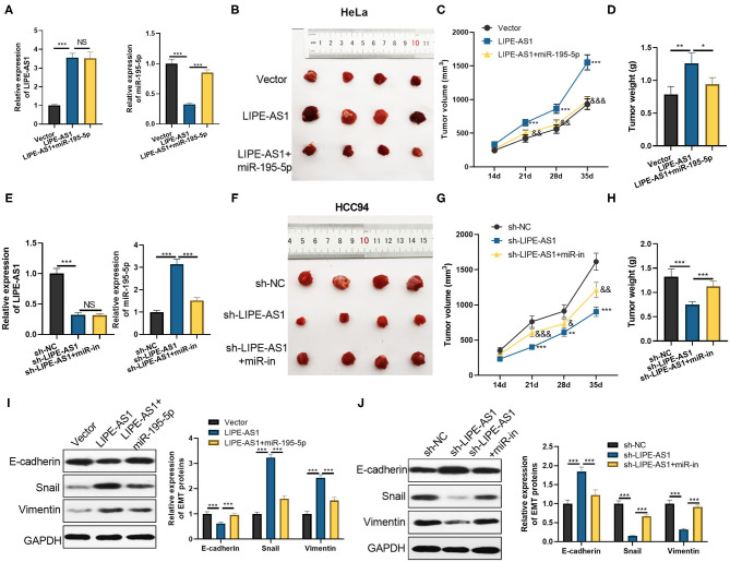
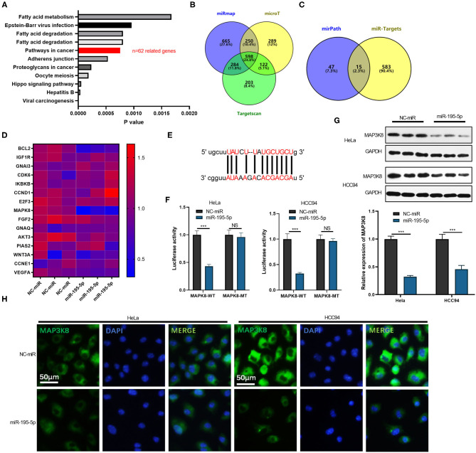
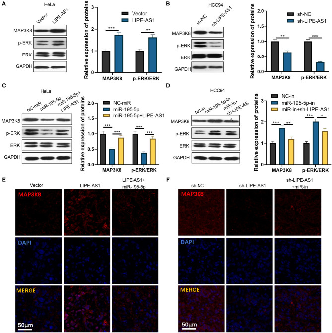
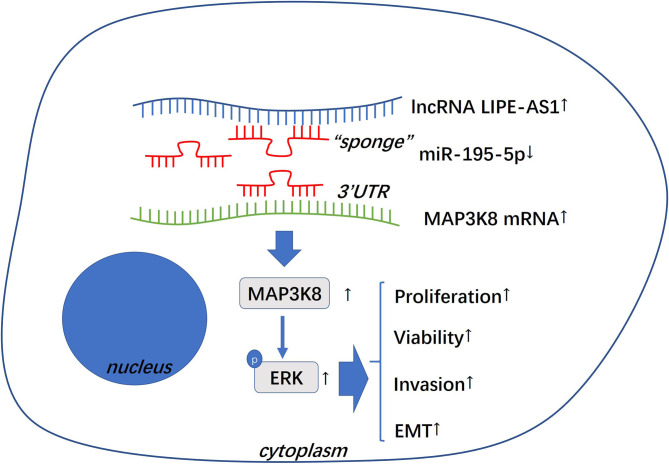
Aims: A growing number of studies have unveiled that long non-coding RNA (lncRNA) is conductive to cervical cancer (CC) development. However, the effect of LIPE-AS1 is remained to be studied in CC. Main Methods: Reverse transcription-polymerase chain reaction (RT-PCR) was employed to measure LIPE-AS1 expression in CC tissues and the adjacent normal tissues. Additionally, we conducted gain- and loss-of functional experiments of LIPE-AS1 and adopted CCK8 assay, BrdU assay, and in vivo tumor formation experiment to test the proliferation of CC cells (HCC94 and HeLa). Besides, the apoptosis, invasion, and epithelial-mesenchymal transformation (EMT) of CC cells were estimated using flow cytometry, transwell assay, and western blot, respectively. Further, LIPE-AS1 downstream targets were analyzed through bioinformatics, and the binding relationships between LIPE-AS1 and miR-195-5p were verified via dual-luciferase activity experiment and RNA Protein Immunoprecipitation (RIP) assay. Moreover, rescue experiments were conducted to confirm the effects of LIPE-AS1 and miR-195-5p in regulating CC development and the expressions of MAPK signaling pathway related proteins were detected by RT-PCR, western blot, and immunofluorescence. Key Findings: LIPE-AS1 was over-expressed in CC tissues (compared to normal adjacent tissues) and was notably related to tumor volume, distant metastasis. Overexpressing LIPE-AS1 accelerated CC cell proliferation, migration and EMT, inhibited apoptosis; while LIPE-AS1 knockdown had the opposite effects. The mechanism studies confirmed that LIPE-AS1 sponges miR-195-5p as a competitive endogenous RNA (ceRNA), which targets the 3'-untranslated region (3'-UTR) of MAP3K8. LIPE-AS1 promoted the expression of MAP3K8 and enhanced ERK1/2 phosphorylation, which were reversed by miR-195-5p. Significance: LIPE-AS1 regulates CC progression through the miR-195-5p/MAPK signaling pathway, providing new hope for CC diagnosis and treatment.

Keywords: LIPE-AS1; MAP3K8; MAPK signaling pathway; cervical cancer; miR-195-5p.


Conflict of interest statement

The authors declare that the research was conducted in the absence of any commercial or financial relationships that could be construed as a potential conflict of interest.

Figures

Figure 1
Figure 1
LIPE-AS1 is increased in cervical cancer and related to worse survival. (A,B) LIPE-AS1 expression in 42 cases of cervical cancer tissues and the adjacent normal tissues were detected by RT-PCR, *P < 0.05, ***P < 0.001. (C) LIPE-AS1 expression in Cervical squamous cell carcinoma and endocervical adenocarcinoma (CESC) was analyzed through the GEPIA database (http://gepia.cancer-pku.cn/). (D) RT-PCR was taken to measure LIPE-AS1 expression in normal cervical epithelial cell lines HUCEC and cervical cancer cell lines (including Hela, MS751, SiHa, CaSki, C-33A, and HCC94). *P < 0.05, ***P < 0.001 vs. HUCEC group.
Figure 2
Figure 2
LIPE-AS1 promotes cervical cancer cell proliferation, invasion, and EMT. LIPE-AS1 over- and low-expressing cell models were constructed in HeLa (A) and HCC94 cells (B), respectively. CCK8 was applied to examine the proliferation of HeLa (C) and HCC94 cells (D). *P < 0.05, **P < 0.01 vs. Vector group or sh-NC group. BrdU method was taken to examine the proliferation of HeLa (E) and HCC94 cells (F). Flow cytometry was performed to test the apoptosis of HeLa (G) and HCC94 cells (H). Transwell method was performed to evaluate the invasion of HeLa (I) and HCC94 cells (J). (K) The expressions of E-cadherin, Snail and Vimentin in HeLa and HCC94 cells were detected by Western blot. *P < 0.05, **P < 0.01, ***P < 0.001. N = 3.
Figure 3
Figure 3
LIPE-AS1 targets miR-195-5p. (A) Through browsing the Starbase database (http://starbase.sysu.edu.cn/), we analyzed the targeted miRNAs of LIPE-AS1 and found that miR-195-5p had complementary bases paired with LIPE-AS1. (B,C) The expression of miR-195-5p in cervical cancer tissues and adjacent normal tissues was measured by RT-PCR (B), and the correlation between LIPE-AS1 and miR-195-5p in cervical cancer tissues was analyzed by linear regression. (D) miR-195-5p expression in human tumor tissues (data from the TCGA database). (E) The relationship between LIPE-AS1-miR-195-5p was verified via dual luciferase activity experiment. (F) The relationship between LIPE-AS1-miR-195-5p was verified by RIP experiments, and the enrichment level of LIPE-AS1 in cell lysate was detected via RT-PCR. NSP > 0.05, ***P < 0.001. (G) RT-PCR was applied to determine miR-195-5p expression in normal cervical epithelial cell line HUCEC and cervical cancer cell lines (including Hela, MS751, SiHa, CaSki, C-33A, and HCC94). **P < 0.01, ***P < 0.001 vs. HUCEC group. (H,I) RT-PCR detected changes in miR-195-5p after silencing or overexpressing LIPE-AS1. ***P < 0.001. N = 3. The arrow shows that miR-195-5p is down-regulated in CESC (Cervical squamous cell carcinoma and endocervical adenocarcinoma) tissues compared with that in the normal tissues.
Figure 4
Figure 4
The function of LIPE-AS1/miR-195-5p in cervical cancer cells. (A) MiR-195-5p mimics and inhibitors were taken to construct miR-195-5p over- and down-expressing cell lines, respectively, and further transfected with LIPE-AS1 over-expressed plasmids or sh-LIPE-AS1 to perform compensation experiments. CCK8 method (B) and BrdU method (C) were employed to detect HeLa and HCC94 cell proliferation. (D) Flow cytometry was performed to test the apoptosis of HeLa and HCC94 cells. (E) Transwell method was carried out to examine HeLa and HCC94 cell invasion. The expressions of E-cadherin, Snail, and Vimentin in HeLa (F) and HCC94 cells (G) were examined by Western blot. **P < 0.01, ***P < 0.001 vs. Vector or sh-NC group. &P < 0.05, &&P < 0.01, &&&P < 0.001 vs. LIPE-AS1 group or sh-LIPE -AS1 group. N = 3.
Figure 5
Figure 5
The function of LIPE-AS1/miR-195-5p in cervical cancer cells in vivo. LIPE-AS1 plasmid or sh-LIPE-AS1 were used to construct cell models of LIPE-AS1 overexpression and knockdown, respectively, and further transfected with miR-195-5p mimic or inhibitor. The nude mouse tumor formation model was constructed with stably transfected cells. The expressions of LIPE-AS1 and miR-195-5p in HeLa cells (A) and HCC94 cells (E) were examined by RT-PCR, respectively. (B,F) Nude mice were sacrificed in the fifth week, and tumor tissue was removed. (C,G) Tumor volume in nude mice; (D,H) tumor mass at the fifth week; (I,J) western blot was taken to determine the expressions of E-cadherin, Snail, and Vimentin in tumor tissues. NSP > 0.05, *P < 0.05, **P < 0.01, ***P < 0.001. &P < 0.05, &&P < 0.01, &&&P < 0.001 vs. LIPE-AS1 group or sh-LIPE-AS1 group. N = 4.
Figure 6
Figure 6
MiR-195-5p targets MAP3K8. (A) The mirpath database (http://snf-515788.vm.okeanos.grnet.gr/index.php?r=mirpath) was used to predict the signal pathways regulated by miR-195-5p. (B) Through mirmap (http://mirnamap.mbc.nctu.edu.tw/), microT (https://omictools.com/diana-microt-cds-tool), and Targetscan (http://www.targetscan.org/vert_72/), miR-195-5p's targeting molecule was predicted. (C) The miR-195-5p related molecules in mirpath, mirmap, microT, and Targetscan databases were analyzed by Venn diagram. (D) RT-PCR was performed to measure tumor gene expression after miR-195-5p overexpression. (E) Base pairing between miR-195-5p and MAPK8. (F) Dual luciferase activity experiments were applied to verify the relationship between miR-195-5p and MAPK8. (G) Western blot was taken to evaluate the protein level of MAP3K8 in over-expressing miR-195-5p and low-expressing miR-195-5p cells. (H) Cell immunofluorescence was taken to evaluate the protein level of MAP3K8 in over-expressing miR-195-5p and low-expressing miR-195-5p cells. NSP > 0.05, ***P < 0.001. N = 3.
Figure 7
Figure 7
The effect of LIPE-AS1/miR-195-5p on the MAPK/ERK pathway. LIPE-AS1 over- and low-expressing cell models were constructed in HeLa (A) and HCC94 cells (B), respectively. Western blot was used to measure MAP3K8 and ERK protein expression in the cells. (C,D) Transfection of cervical cancer cells with miR-195-5p mimics and inhibitors, LIPE-AS1 over-expressed plasmid or sh-LIPE-AS1. Western blot was taken to measure the protein expressions of MAP3K8 and ERK in cells. (E,F) Tissue immunofluorescence was carried out to detect MAP3K8 in the tumor tissues (the grouping was the same as Figure 5). *P < 0.05, **P < 0.01, ***P < 0.001. N = 3.
Figure 8
Figure 8
The mechanism diagram. During the progression of cervical cancer (CC), LIPE-AS1 is upregulated and inhibits the expression of miR-195-5p, which targets at the 3′UTR of MAP3K8 mRNA. Low level of miR-195-5p leads to overexpressed MAP3K8 thus enhancing the phosphorylation of ERK. The activation of MAPK/ERK pathway increases the proliferation, viability, invasion, and EMT of CC cells, therefore promoting the development of CC.

Similar articles

Cited by

References

    1. Ãukić A, Lulić L, Thomas M, Skelin J, Bennett Saidu NE, Grce M, et al. . HPV oncoproteins and the ubiquitin proteasome system: a signature of malignancy? Pathogens. (2020) 9:133. 10.3390/pathogens9020133 - DOI - PMC - PubMed
    1. Łaniewski P, Ilhan ZE, Herbst-Kralovetz MM. The microbiome and gynaecological cancer development, prevention and therapy. Nat Rev Urol. (2020) 17:232–50. 10.1038/s41585-020-0286-z - DOI - PMC - PubMed
    1. Aalijahan H, Ghorbian S. Long non-coding RNAs and cervical cancer. Exp Mol Pathol. (2019) 106:7–16. 10.1016/j.yexmp.2018.11.010 - DOI - PubMed
    1. Wang AH, Jin CH, Cui GY, Li HY, Wang Y, Yu JJ, et al. . MIR210HG promotes cell proliferation and invasion by regulating miR-503-5p/TRAF4 axis in cervical cancer. Aging. (2020) 12:3205–17. 10.18632/aging.102799 - DOI - PMC - PubMed
    1. Li R, Liu J, Qi J. Knockdown of long non-coding RNA CCAT1 suppresses proliferation and EMT of human cervical cancer cell lines by down-regulating Runx2. Exp Mol Pathol. (2020) 113:104380. 10.1016/j.yexmp.2020.104380 - DOI - PubMed

Publication types

LinkOut - more resources