

Phagocytosis by Peripheral Glia: Importance for Nervous System Functions and Implications in Injury and Disease
- PMID: 33898462
- PMCID: PMC8060502
- DOI: 10.3389/fcell.2021.660259
Phagocytosis by Peripheral Glia: Importance for Nervous System Functions and Implications in Injury and Disease
Abstract
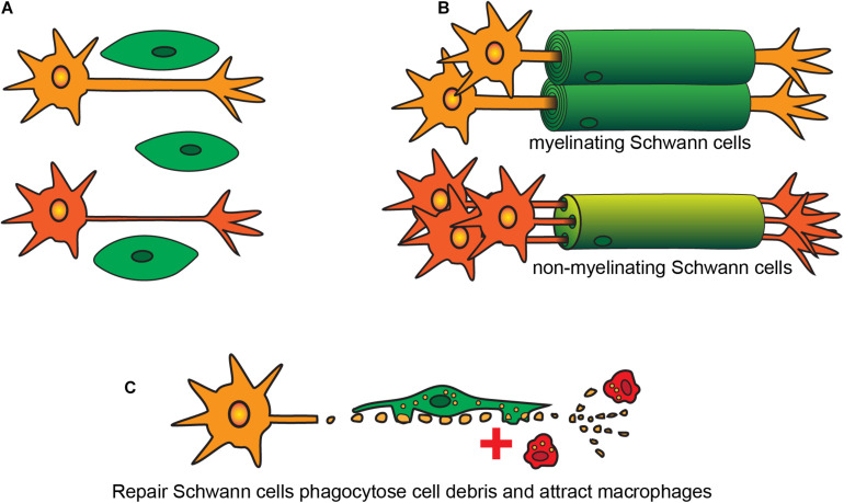
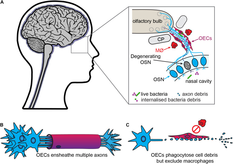
The central nervous system (CNS) has very limited capacity to regenerate after traumatic injury or disease. In contrast, the peripheral nervous system (PNS) has far greater capacity for regeneration. This difference can be partly attributed to variances in glial-mediated functions, such as axon guidance, structural support, secretion of growth factors and phagocytic activity. Due to their growth-promoting characteristic, transplantation of PNS glia has been trialed for neural repair. After peripheral nerve injuries, Schwann cells (SCs, the main PNS glia) phagocytose myelin debris and attract macrophages to the injury site to aid in debris clearance. One peripheral nerve, the olfactory nerve, is unique in that it continuously regenerates throughout life. The olfactory nerve glia, olfactory ensheathing cells (OECs), are the primary phagocytes within this nerve, continuously clearing axonal debris arising from the normal regeneration of the nerve and after injury. In contrast to SCs, OECs do not appear to attract macrophages. SCs and OECs also respond to and phagocytose bacteria, a function likely critical for tackling microbial invasion of the CNS via peripheral nerves. However, phagocytosis is not always effective; inflammation, aging and/or genetic factors may contribute to compromised phagocytic activity. Here, we highlight the diverse roles of SCs and OECs with the focus on their phagocytic activity under physiological and pathological conditions. We also explore why understanding the contribution of peripheral glia phagocytosis may provide us with translational strategies for achieving axonal regeneration of the injured nervous system and potentially for the treatment of certain neurological diseases.
Keywords: Schwann cell; bacteria; cell debris; macrophage; neuropathy; olfactory ensheathing cell.
Copyright © 2021 Nazareth, St John, Murtaza and Ekberg.
Conflict of interest statement
The authors declare that the research was conducted in the absence of any commercial or financial relationships that could be construed as a potential conflict of interest.
Figures
References
-
- Acosta C. C. D., Dias A. A., Rosa T., Batista-Silva L. R., Rosa P. S., Toledo-Pinto T. G., et al. (2018). Pgl i expression in live bacteria allows activation of a cd206/ppar gamma cross-talk that may contribute to successful mycobacterium leprae colonization of peripheral nerves. PLos Pathog. 14:1007151. 10.1371/journal.ppat.1007151 - DOI - PMC - PubMed
-
- Alves L., Lima L. D. M., Maeda E. D. S., Carvalho L., Holy J., Sarno E. N., et al. (2004). Mycobacterium leprae infection of human schwann cells depends on selective host kinases and pathogen-modulated endocytic pathways. Fems Microbiol. Lett. 238 429–437. 10.1016/j.femsle.2004.08.007 - DOI - PubMed
Publication types
LinkOut - more resources
Full Text Sources
Other Literature Sources