

Molecular Landscapes and Models of Acute Erythroleukemia
- PMID: 33898929
- PMCID: PMC8061683
- DOI: 10.1097/HS9.0000000000000558
Molecular Landscapes and Models of Acute Erythroleukemia
Abstract
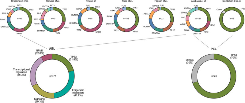
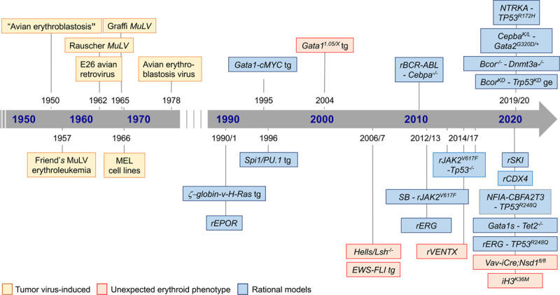
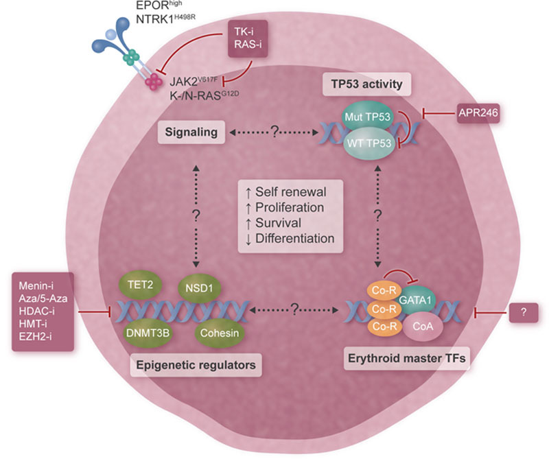
Malignancies of the erythroid lineage are rare but aggressive diseases. Notably, the first insights into their biology emerged over half a century ago from avian and murine tumor viruses-induced erythroleukemia models providing the rationale for several transgenic mouse models that unraveled the transforming potential of signaling effectors and transcription factors in the erythroid lineage. More recently, genetic roadmaps have fueled efforts to establish models that are based on the epigenomic lesions observed in patients with erythroid malignancies. These models, together with often unexpected erythroid phenotypes in genetically modified mice, provided further insights into the molecular mechanisms of disease initiation and maintenance. Here, we review how the increasing knowledge of human erythroleukemia genetics combined with those from various mouse models indicate that the pathogenesis of the disease is based on the interplay between signaling mutations, impaired TP53 function, and altered chromatin organization. These alterations lead to aberrant activity of erythroid transcriptional master regulators like GATA1, indicating that erythroleukemia will most likely require combinatorial targeting for efficient therapeutic interventions.
Copyright © 2021 the Author(s). Published by Wolters Kluwer Health, Inc. on behalf of the European Hematology Association.
Figures
Similar articles
-
Erythroleukemia-historical perspectives and recent advances in diagnosis and management.Blood Rev. 2018 Mar;32(2):96-105. doi: 10.1016/j.blre.2017.09.002. Epub 2017 Sep 18. Blood Rev. 2018. PMID: 28965757 Free PMC article. Review.
-
Expression of c-MYC under the control of GATA-1 regulatory sequences causes erythroleukemia in transgenic mice.J Exp Med. 1995 May 1;181(5):1603-13. doi: 10.1084/jem.181.5.1603. J Exp Med. 1995. PMID: 7722440 Free PMC article.
-
Induction of erythrocyte protein 4.2 gene expression during differentiation of murine erythroleukemia cells.Genomics. 1999 Jul 1;59(1):6-17. doi: 10.1006/geno.1999.5846. Genomics. 1999. PMID: 10395794
-
Pluripotent stem cells reveal erythroid-specific activities of the GATA1 N-terminus.J Clin Invest. 2015 Mar 2;125(3):993-1005. doi: 10.1172/JCI75714. Epub 2015 Jan 26. J Clin Invest. 2015. PMID: 25621499 Free PMC article.
-
Revisiting erythroleukemia.Curr Opin Hematol. 2017 Mar;24(2):146-151. doi: 10.1097/MOH.0000000000000314. Curr Opin Hematol. 2017. PMID: 27875373 Review.
Cited by
-
Acute Erythroid Leukemia: From Molecular Biology to Clinical Outcomes.Int J Mol Sci. 2024 Jun 6;25(11):6256. doi: 10.3390/ijms25116256. Int J Mol Sci. 2024. PMID: 38892446 Free PMC article. Review.
-
HDAC7 is a potential therapeutic target in acute erythroid leukemia.Leukemia. 2024 Dec;38(12):2614-2627. doi: 10.1038/s41375-024-02394-5. Epub 2024 Sep 15. Leukemia. 2024. PMID: 39277669 Free PMC article.
-
Epigenetic Regulation of Erythropoiesis: From Developmental Programs to Therapeutic Targets.Int J Mol Sci. 2025 Jun 30;26(13):6342. doi: 10.3390/ijms26136342. Int J Mol Sci. 2025. PMID: 40650116 Free PMC article. Review.
-
Erythroid Cell Research: 3D Chromatin, Transcription Factors and Beyond.Int J Mol Sci. 2022 May 30;23(11):6149. doi: 10.3390/ijms23116149. Int J Mol Sci. 2022. PMID: 35682828 Free PMC article. Review.
References
-
- Bain BJ. Di Guglielmo and his syndromes. Br J Haematol. 2003;120:939–943. - PubMed
-
- Bennett JM, Catovsky D, Daniel MT, et al. . Proposals for the classification of the acute leukaemias. French-American-British (FAB) Co-operative Group. Br J Haematol. 1976;33:451–458. - PubMed
-
- Vardiman JW, Thiele J, Arber DA, et al. . The 2008 revision of the World Health Organization (WHO) classification of myeloid neoplasms and acute leukemia: rationale and important changes. Blood. 2009;114:937–951. - PubMed
-
- Arber DA, Orazi A, Hasserjian R, et al. . The 2016 revision to the World Health Organization classification of myeloid neoplasms and acute leukemia. Blood. 2016;127:2391–2405. - PubMed
Publication types
LinkOut - more resources
Full Text Sources
Other Literature Sources
Research Materials
Miscellaneous