

Deep Learning-Enabled Label-Free On-Chip Detection and Selective Extraction of Cell Aggregate-Laden Hydrogel Microcapsules
- PMID: 33899299
- PMCID: PMC8203426
- DOI: 10.1002/smll.202100491
Deep Learning-Enabled Label-Free On-Chip Detection and Selective Extraction of Cell Aggregate-Laden Hydrogel Microcapsules
Erratum in
-
Deep Learning-Enabled Label-Free On-Chip Detection and Selective Extraction of Cell Aggregate-Laden Hydrogel Microcapsules.Small. 2021 Jun;17(24):e2102868. doi: 10.1002/smll.202102868. Small. 2021. PMID: 34145733 No abstract available.
Abstract
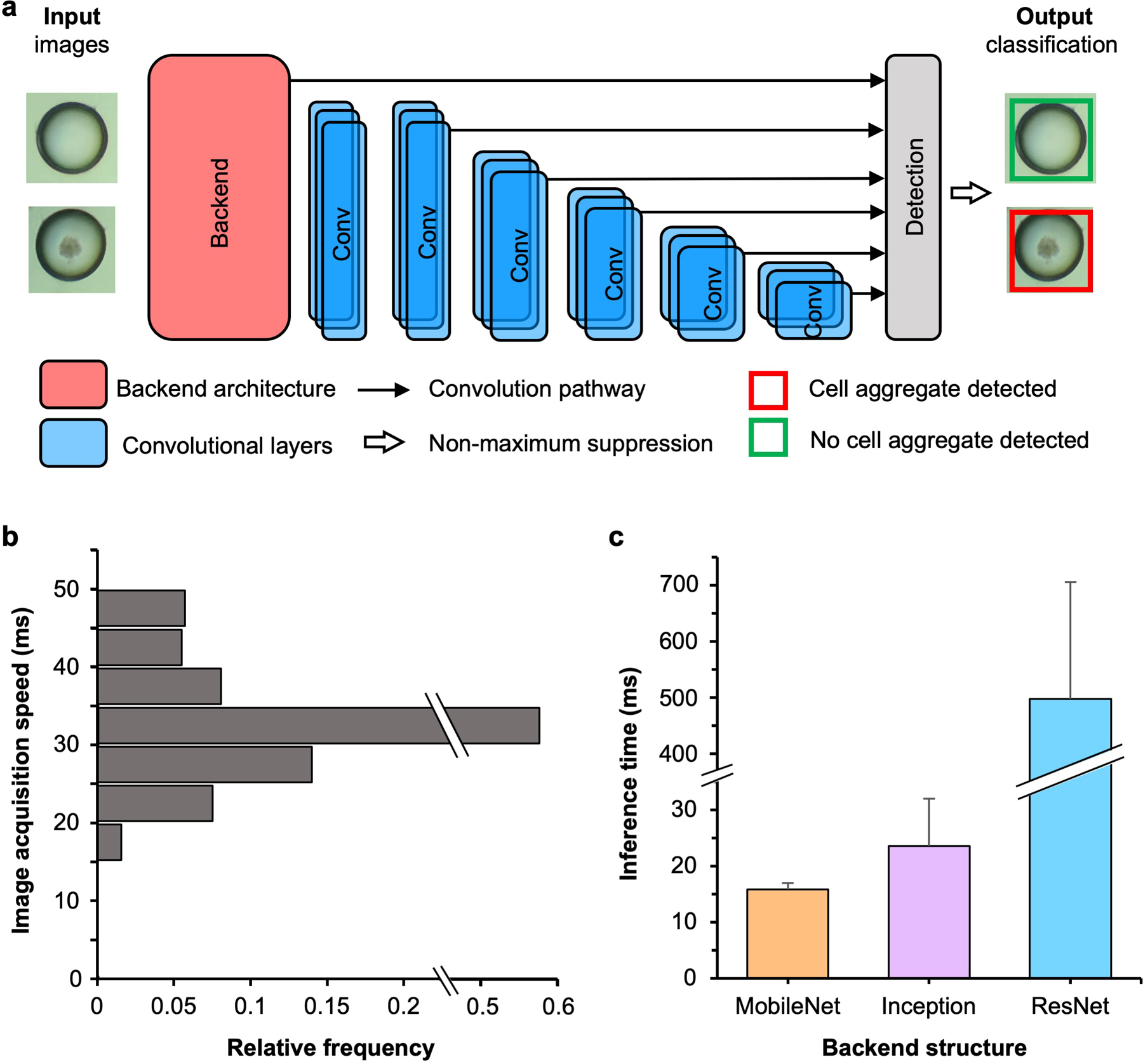
Microfluidic encapsulation of cells/tissues in hydrogel microcapsules has attracted tremendous attention in the burgeoning field of cell-based medicine. However, when encapsulating rare cells and tissues (e.g., pancreatic islets and ovarian follicles), the majority of the resultant hydrogel microcapsules are empty and should be excluded from the sample. Furthermore, the cell-laden hydrogel microcapsules are usually suspended in an oil phase after microfluidic generation, while the microencapsulated cells require an aqueous phase for further culture/transplantation and long-term suspension in oil may compromise the cells/tissues. Thus, real-time on-chip selective extraction of cell-laden hydrogel microcapsules from oil into aqueous phase is crucial to the further use of the microencapsulated cells/tissues. Contemporary extraction methods either require labeling of cells for their identification along with an expensive detection system or have a low extraction purity (<≈30%). Here, a deep learning-enabled approach for label-free detection and selective extraction of cell-laden microcapsules with high efficiency of detection (≈100%) and extraction (≈97%), high purity of extraction (≈90%), and high cell viability (>95%) is reported. The utilization of deep learning to dynamically analyze images in real time for label-free detection and on-chip selective extraction of cell-laden hydrogel microcapsules is unique and may be valuable to advance the emerging cell-based medicine.
Keywords: cell microencapsulation; hydrogel; machine learning; microfluidic; transplantation.
© 2021 Wiley-VCH GmbH.
Conflict of interest statement
Competing and financial interests
The authors declare no competing financial interests.
Figures


Similar articles
-
Label-Free On-Chip Selective Extraction of Cell-Aggregate-Laden Microcapsules from Oil into Aqueous Solution with Optical Sensor and Dielectrophoresis.ACS Sens. 2018 Feb 23;3(2):410-417. doi: 10.1021/acssensors.7b00834. Epub 2018 Jan 24. ACS Sens. 2018. PMID: 29299919 Free PMC article.
-
Generation and manipulation of hydrogel microcapsules by droplet-based microfluidics for mammalian cell culture.Lab Chip. 2017 May 31;17(11):1913-1932. doi: 10.1039/c7lc00262a. Lab Chip. 2017. PMID: 28509918 Free PMC article. Review.
-
Stiffness-Independent Highly Efficient On-Chip Extraction of Cell-Laden Hydrogel Microcapsules from Oil Emulsion into Aqueous Solution by Dielectrophoresis.Small. 2015 Oct 28;11(40):5369-74. doi: 10.1002/smll.201501388. Epub 2015 Aug 21. Small. 2015. PMID: 26297051 Free PMC article.
-
Deep Learning-Enabled Label-Free On-Chip Detection and Selective Extraction of Cell Aggregate-Laden Hydrogel Microcapsules.Small. 2021 Jun;17(24):e2102868. doi: 10.1002/smll.202102868. Small. 2021. PMID: 34145733 No abstract available.
-
Microfluidic fabrication of microengineered hydrogels and their application in tissue engineering.Lab Chip. 2012 Jan 7;12(1):45-59. doi: 10.1039/c1lc20859d. Epub 2011 Nov 21. Lab Chip. 2012. PMID: 22105780 Review.
Cited by
-
Primary Human Pancreatic Cancer Cells Cultivation in Microfluidic Hydrogel Microcapsules for Drug Evaluation.Adv Sci (Weinh). 2023 Apr;10(12):e2206004. doi: 10.1002/advs.202206004. Epub 2023 Feb 19. Adv Sci (Weinh). 2023. PMID: 36808707 Free PMC article.
-
A Microfluidic Approach for Probing Heterogeneity in Cytotoxic T-Cells by Cell Pairing in Hydrogel Droplets.Micromachines (Basel). 2022 Nov 4;13(11):1910. doi: 10.3390/mi13111910. Micromachines (Basel). 2022. PMID: 36363930 Free PMC article.
-
Microencapsulation and nanowarming enables vitrification cryopreservation of mouse preantral follicles.Nat Commun. 2022 Dec 15;13(1):7515. doi: 10.1038/s41467-022-34549-2. Nat Commun. 2022. PMID: 36522314 Free PMC article.
-
Towards single cell encapsulation for precision biology and medicine.Adv Drug Deliv Rev. 2023 Oct;201:115010. doi: 10.1016/j.addr.2023.115010. Epub 2023 Jul 16. Adv Drug Deliv Rev. 2023. PMID: 37454931 Free PMC article. Review.
-
Artificial Intelligence-Empowered Automated Double Emulsion Droplet Library Generation.Small. 2025 May;21(18):e2412099. doi: 10.1002/smll.202412099. Epub 2025 Mar 25. Small. 2025. PMID: 40130763 Free PMC article.
References
-
- Zhang J, Coulston RJ, Jones ST, Geng J, Scherman OA, Abell C, Science (80-. ) 2012, 335, 690. - PubMed
-
- Vegas AJ, Veiseh O, Gürtler M, Millman JR, Pagliuca FW, Bader AR, Doloff JC, Li J, Chen M, Olejnik K, Tam HH, Jhunjhunwala S, Langan E, Aresta-Dasilva S, Gandham S, McGarrigle JJ, Bochenek MA, Hollister-Lock J, Oberholzer J, Greiner DL, Weir GC, Melton DA, Langer R, Anderson DG, Nat. Med 2016, 22, 306. - PMC - PubMed
Publication types
MeSH terms
Substances
Grants and funding
LinkOut - more resources
Full Text Sources
