Review
. 2021 Aug;246(16):1816-1828.
doi: 10.1177/15353702211009146. Epub 2021 Apr 25.

Bioengineering approaches to mature induced pluripotent stem cell-derived atrial cardiomyocytes to model atrial fibrillation

Affiliations
Review

Bioengineering approaches to mature induced pluripotent stem cell-derived atrial cardiomyocytes to model atrial fibrillation

Olivia T Ly et al. Exp Biol Med (Maywood). 2021 Aug.

Abstract

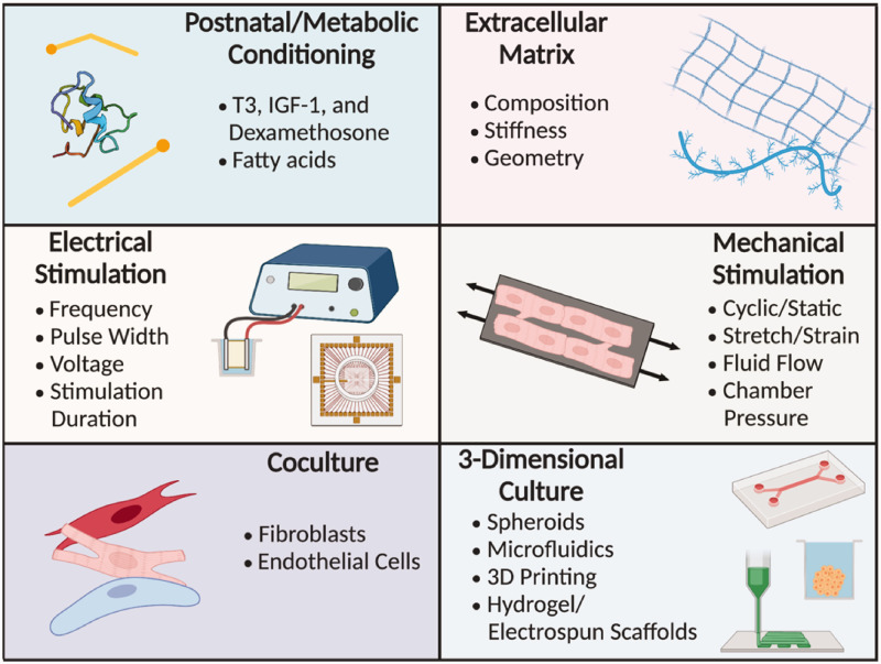
Induced pluripotent stem cells (iPSCs) serve as a robust platform to model several human arrhythmia syndromes including atrial fibrillation (AF). However, the structural, molecular, functional, and electrophysiological parameters of patient-specific iPSC-derived atrial cardiomyocytes (iPSC-aCMs) do not fully recapitulate the mature phenotype of their human adult counterparts. The use of physiologically inspired microenvironmental cues, such as postnatal factors, metabolic conditioning, extracellular matrix (ECM) modulation, electrical and mechanical stimulation, co-culture with non-parenchymal cells, and 3D culture techniques can help mimic natural atrial development and induce a more mature adult phenotype in iPSC-aCMs. Such advances will not only elucidate the underlying pathophysiological mechanisms of AF, but also identify and assess novel mechanism-based therapies towards supporting a more 'personalized' (i.e. patient-specific) approach to pharmacologic therapy of AF.

Keywords: Atrial fibrillation; disease modeling; human-induced pluripotent stem cell-derived atrial cardiomyocytes; maturation; pharmacologic response.


Conflict of interest statement

DECLARATION OF CONFLICTING INTERESTS: The author(s) declared no potential conflicts of interest with respect to the research, authorship, and/or publication of this article.

Figures

Figure 1.
Figure 1.
Differentiation of iPSCs to atrial cardiomyocytes: (a) Protocol for differentiation of iPSCs into atrial cardiomyocytes. iPSCs are differentiated into cardiomyocytes using a commercially available cardiomyocyte differentiation medium for 5 days, then incubated with 1 μM retinoic acid (RA) or DMSO (vehicle control or CT) for another 5 days to enrich iPSC-aCMs, and the iPSC-aCMs are further enriched via glucose starvation for 5 more days, for a total of 15 days. (b) Immunostaining showing the protein expression of pan-CM marker cardiac cTnT and atrial marker Kv1.5 in hiPSC-CMs at day 10 comparing RA treated cells to CT cells. (c) qRT-PCR of ventricular marker, MYH7, in RA-treated and CT cells and atrial markers, KCNJ3 and KCNA5, in RA-treated and CT cells at day 30. Figure adapted from Argenziano et al. (A color version of this figure is available in the online journal.)
Figure 2.
Figure 2.
Atrial action potential: the action potential is initiated by a depolarization (Phase 0) caused by a rapid influx of Na+ ions, followed by a transient repolarization (Phase 1) mediated by an efflux of K+ balanced with an influx of Ca2+. This is followed by a plateau phase (Phase 2) maintained by massive influx of Ca2+ through calcium-induced calcium release from the sarcoplasmic reticulum, and efflux of K+ through the quickly activating but slower inactivating ultrarapid delayed rectifier, followed by a late repolarization phase (Phase 3) primarily induced by dissipation of Ca2+ and K+ efflux through activation of several K+ channels, before returning to the resting state (Phase 4). (A color version of this figure is available in the online journal.)
Figure 3.
Figure 3.
Methods for iPSC-derived atrial cardiomyocyte maturation: postnatal and metabolic conditioning, extracellular matrix, electrical stimulation, mechanical stimulation, co-culture, and three-dimensional culture have all been utilized to increase iPSC-derived cardiomyocyte maturity in culture. Figure created using BioRender.com. (A color version of this figure is available in the online journal.)
Figure 4.
Figure 4.
Bioengineered models of cardiomyocytes and cardiac tissue: (a) Treatment of iPSC-CMs with postnatal factors triiodothyronine (T3), insulin-like growth factor-1 (IGF-1), and dexamethasone (Dex) leads to increased cardiomyocyte size and sarcomere alignment. Figure adapted from Birket et al. (b) Representative traces for control and fatty acid (FA)-treated hPSC-CMs responding to the ATP synthase inhibitor oligomycin, the respiratory uncoupler FCCP, and the respiratory chain blockers, rotenone and antimycin A. Higher maximal oxygen consumption rate (OCR) was seen in FA-treated versus control (CNTL) cells. Upstroke velocity (right) was also increased with FA treatment. Figure adapted from Yang et al. (c) Micropatterned cardiomyocytes led to alignment of cardiomyocytes and their actin filaments. Figure adapted from Salick et al. (d) Electrical stimulation promoted improvement in Ca2+ handling properties as evident by non-stimulated control cells not responding to caffeine while stimulated cells responded to caffeine by releasing more calcium ions. Fluorescence recording (right) of calcium transients before and after administration of caffeine (arrow) in cells exposed to 6-Hz electrical stimulation. Figure adapted from Sun and Nunes. (e) Mechanical stimulation of iPSC-CMs led to an increase in Connexin 43 (Cx43 and Cnx43) formation compared with static controls. Figure adapted from Mihic et al. (f) Three-dimensional culture (N-cadherin (red) and EH-myomesin (green)) as well as co-culture with cardiac fibroblasts (CF) lead to increases in APD and amplitude (AMP). Figure adapted from Beauchamp et al. (A color version of this figure is available in the online journal.)

References

    1. Chugh SS, Havmoeller R, Narayanan K, Singh D, Rienstra M, Benjamin EJ, Gillum RF, Kim Y-H, McAnulty JH, Zheng Z-J, Forouzanfar MH, Naghavi M, Mensah GA, Ezzati M, Murray CJL. Worldwide epidemiology of atrial fibrillation. Circulation 2014; 129:837–47 - PMC - PubMed
    1. Stewart S, Murphy NF, Murphy N, Walker A, McGuire A, McMurray JJV. Cost of an emerging epidemic: an economic analysis of atrial fibrillation in the UK. Heart 2004; 90:286–92 - PMC - PubMed
    1. Miyasaka Y, Barnes ME, Gersh BJ, Cha SS, Bailey KR, Abhayaratna WP, Seward JB, Tsang TSM. Secular trends in incidence of atrial fibrillation in Olmsted county, Minnesota, 1980 to 2000, and implications on the projections for future prevalence. Circulation 2006; 114:119–25 - PubMed
    1. Lloyd-Jones DM, Wang TJ, Leip EP, Larson MG, Levy D, Vasan RS, D’Agostino RB, Massaro JM, Beiser A, Wolf PA, Benjamin EJ. Lifetime risk for development of atrial fibrillation: the Framingham heart study. Circulation 2004; 110:1042–6 - PubMed
    1. Darbar D, Roden DM. Genetic mechanisms of atrial fibrillation: impact on response to treatment. Nat Rev Cardiol 2013; 10:317–29 - PMC - PubMed

Publication types

MeSH terms