

Hemodynamic and oxygen-metabolic responses of the awake mouse brain to hypercapnia revealed by multi-parametric photoacoustic microscopy
- PMID: 33899557
- PMCID: PMC8504963
- DOI: 10.1177/0271678X211010352
Hemodynamic and oxygen-metabolic responses of the awake mouse brain to hypercapnia revealed by multi-parametric photoacoustic microscopy
Abstract
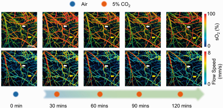
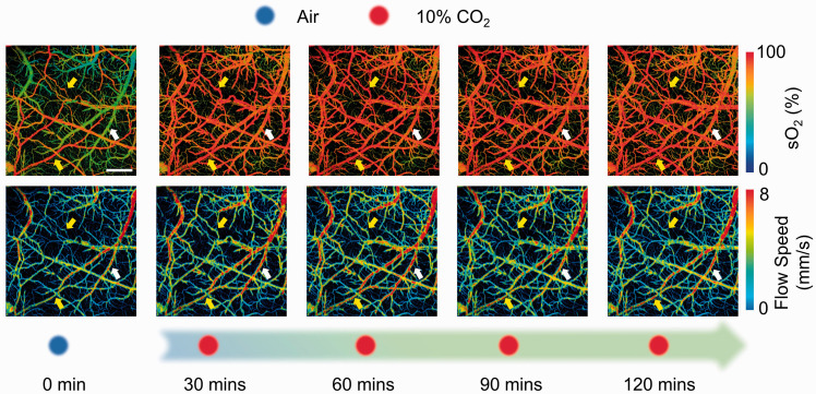
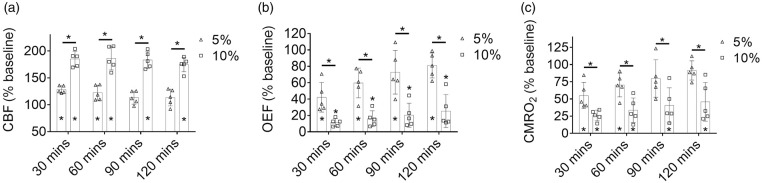
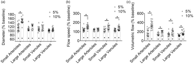
A widely used cerebrovascular stimulus and common pathophysiologic condition, hypercapnia is of great interest in brain research. However, it remains controversial how hypercapnia affects brain hemodynamics and energy metabolism. By using multi-parametric photoacoustic microscopy, the multifaceted responses of the awake mouse brain to different levels of hypercapnia are investigated. Our results show significant and vessel type-dependent increases of the vessel diameter and blood flow in response to the hypercapnic challenges, along with a decrease in oxygen extraction fraction due to elevated venous blood oxygenation. Interestingly, the increased blood flow and decreased oxygen extraction are not commensurate with each other, which leads to reduced cerebral oxygen metabolism. Further, time-lapse imaging over 2-hour chronic hypercapnic challenges reveals that the structural, functional, and metabolic changes induced by severe hypercapnia (10% CO2) are not only more pronounced but more enduring than those induced by mild hypercapnia (5% CO2), indicating that the extent of brain's compensatory response to chronic hypercapnia is inversely related to the severity of the challenge. Offering quantitative, dynamic, and CO2 level-dependent insights into the hemodynamic and metabolic responses of the brain to hypercapnia, these findings might provide useful guidance to the application of hypercapnia in brain research.
Keywords: Photoacoustic microscopy; hemodynamics; hypercapnia; oxygen metabolism; vascular response.
Conflict of interest statement
Figures

References
-
- Ito H, Kanno I, Ibaraki M, et al. Changes in human cerebral blood flow and cerebral blood volume during hypercapnia and hypocapnia measured by positron emission tomography. J Cereb Blood Flow Metab 2003; 23: 665–670. - PubMed
-
- Bishop CC, Powell S, Rutt D, et al. Transcranial Doppler measurement of middle cerebral artery blood flow velocity: a validation study. Stroke 1986; 17: 913–915. - PubMed
-
- Jones M, Berwick J, Hewson-Stoate N, et al. The effect of hypercapnia on the neural and hemodynamic responses to somatosensory stimulation. Neuroimage 2005; 27: 609–623. - PubMed
Publication types
MeSH terms
Substances
Grants and funding
LinkOut - more resources
Full Text Sources
Other Literature Sources
