. 2021 Dec;15(1):73-88.
doi: 10.1080/19336934.2021.1915683.

Characterizing a gene expression toolkit for eye- and photoreceptor-specific expression in Drosophila

Affiliations

Characterizing a gene expression toolkit for eye- and photoreceptor-specific expression in Drosophila

Spencer E Escobedo et al. Fly (Austin). 2021 Dec.

Abstract

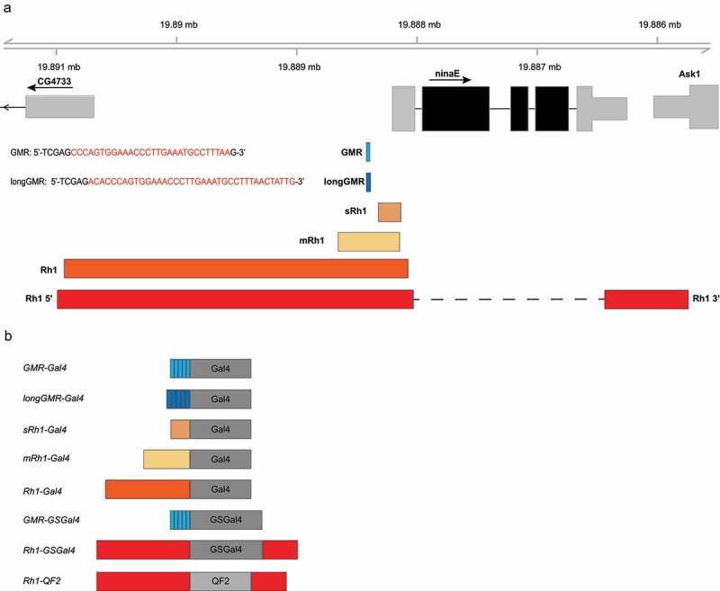
Binary expression systems are a powerful tool for tissue- and cell-specific research. Many of the currently available Drosophila eye-specific drivers have not been systematically characterized for their expression level and cell-type specificity in the adult eye or during development. Here, we used a luciferase reporter to measure expression levels of different drivers in the adult Drosophila eye, and characterized the cell type-specificity of each driver using a fluorescent reporter in live 10-day-old adult males. We also further characterized the expression pattern of these drivers in various developmental stages. We compared several Gal4 drivers from the Bloomington Drosophila Stock Center (BDSC) including GMR-Gal4, longGMR-Gal4 and Rh1-Gal4 with newly developed Gal4 and QF2 drivers that are specific to different cell types in the adult eye. In addition, we generated drug-inducible Rh1-GSGal4 lines and compared their induced expression with an available GMR-GSGal4 line. Although both lines had significant induction of gene expression measured by luciferase activity, Rh1-GSGal4 was expressed at levels below the detection of the fluorescent reporter by confocal microscopy, while GMR-GSGal4 showed substantial reporter expression in the absence of drug by microscopy. Overall, our study systematically characterizes and compares a large toolkit of eye- and photoreceptor-specific drivers, while also uncovering some of the limitations of currently available expression systems in the adult eye.

Keywords: Drosophila; GMR; Gal4 expression system; Geneswitch Gal4; QF2 expression system; Rh1; photoreceptor.


Conflict of interest statement

No potential conflict of interest was reported by the authors.

Figures

Figure 1.
Figure 1.
Overview of the ninaE regulatory regions used in the Gal4 and QF2 driver constructs. a) Schematic of the ninaE locus on chromosome 3 (3 R:19,888,206) showing relative positions of the regulatory elements used in each of the driver constructs. Inset text shows the sequences of the Glass transcription factor binding site in the ninaE promoter that were pentamerized to generate the GMR or longGMR sequences. b) The different regulatory elements described in panel A drive expression of the Gal4, GSGal4 or QF2 drivers. The sRh1-Gal4, mRh1-Gal4, Rh1-GSGal4 and Rh1-QF2 drivers were generated in this study, and compared with the Rh1-Gal4, GMR-Gal4 and longGMR-Gal4 drivers available from the Bloomington Drosophila Stock Center
Figure 2.
Figure 2.
Rh1 drivers are expressed specifically in adult photoreceptors, whereas GMR, longGMR, sRh1 and mRh1 drivers are expressed throughout the adult eye. The indicated Gal4 and QF2 driver lines were crossed to UAS-mCherry or 10XQUAS-QUAS-mCherry flies, respectively, and confocal microscopy was conducted on 10-day-old live adult male progeny. Representative images for each driver are shown (n = 3). Inset images show a magnified section of the eye. (a) Schematic depicting the location of cells in each ommatidium at a similar focal plane to that shown in each image. mCherry expression pattern for the (b) GMR-Gal4, longGMR-Gal4, sRh1-Gal4, mRh1-Gal4, and Rh1-Gal4 drivers, relative to the UAS-mCherry control; (c) Rh1-QF2 drivers and QUAS-mCherry control; (d) GMR-GSGal4 + drug versus vehicle only control. Open and closed arrow heads point to interommatidial cells (IOCs) and R cells, respectively. Scale bar, 50 µm
Figure 3.
Figure 3.
GMR, longGMR, sRh1 and mRh1 drivers are expressed in embryonic tissue, whereas Rh1 drivers are not. The indicated GAL4 and QF2 driver lines were crossed to UAS-mCherry or QUAS-mCherry flies, respectively, and fluorescent microscopy was conducted on late-stage embryos. (a, b) mCherry and (A’, B’) merged mCherry, DAPI and transmitted light (TL) images for (A, A’) GMR-Gal4, longGMR-Gal4, sRh1-Gal4, mRh1-Gal4, Rh1-Gal4, UAS-mCherry control and (B, B’) Rh1-QF2 drivers and QUAS-mCherry control. Abbreviations for labelled tissues are as follows: (CNS) central nervous system; (VNC) ventral nerve cord; (PN) peripheral nervous system; (BO) Bolwig’s organ; (RG) ring gland. Scale bar, 100 µm
Figure 4.
Figure 4.
GMR, longGMR, sRh1 and mRh1 drivers are expressed in various larval tissues, whereas Rh1 drivers have no detectable expression. The indicated GAL4 and QF2 driver lines were crossed to UAS-mCherry or QUAS-mCherry flies, respectively, and fluorescent microscopy was conducted on wandering third instar larvae. (a) mCherry and (A’) merged mCherry, DAPI and transmitted light (TL) images for (A) UAS-mCherry control, GMR-Gal4, longGMR-Gal4, sRh1-Gal4, mRh1-Gal4, Rh1-Gal4, QUAS-mCherry control, and Rh1-QF2 drivers. Abbreviations for labelled tissues are as follows: (VNC) ventral nerve cord; (EAD) eye antennal disc; (SG) salivary glands; (t) tracheal system; Scale bar, 500 µm
Figure 5.
Figure 5.
GMR, longGMR and Rh1 drivers are expressed eye-specifically, while sRh1 and mRh1 are expressed elsewhere in the late stage pupal head and brain. The indicated GAL4 and QF2 driver lines were crossed to UAS-mCherry or QUAS-mCherry flies, respectively, and fluorescent microscopy was conducted on dissected late stage pupal heads. (a, b) mCherry and (A’, B’) merged mCherry and transmitted light (TL) images for (A, A’) GMR-Gal4, longGMR-Gal4, sRh1-Gal4, mRh1-Gal4, Rh1-Gal4, UAS-mCherry control, and (B, B’) Rh1-QF2 drivers and QUAS-mCherry control. Abbreviations for labelled tissues are as follows: (CB) central brain; (l) lamina; (a) antenna; (MP) mouth parts. Scale bar, 100 µm
Figure 6.
Figure 6.
Gal4 drivers expressed in multiple cell types induce higher expression levels relative to photoreceptor-specific Gal4 or QF2 drivers. The indicated eye- or photoreceptor-specific driver lines were crossed to flies containing UAS-Luc (QUAS-Luc for QF2 drivers), and luciferase activity was assayed in heads from adult male progeny expressing UAS-Luc under control of the indicated driver at 10 days post-eclosion. Data are shown as bar plots of means with individual biological replicates overlaid as dots (n = 4, 2 heads/experiment). Luciferase activity for the (a) GMR-Gal4, longGMR-Gal4; (b) Rh1-Gal4, sRh1-Gal4, and mRh1-Gal4 compared to UAS-Luc alone; (c) photoreceptor-specific Rh1-QF2 drivers on chromosomes 2 or 3, compared to QUAS-Luc alone
Figure 7.
Figure 7.
Gene Switch drivers induce gene expression weakly in the adult eye. (a) GMR-GSGal4 flies were crossed to UAS-Luc flies and seven-day old adult progeny were fed food supplemented with RU486 or vehicle only for 24, 48, 72 hours followed by luciferase activity assays on heads to establish maximum GSGal4 induction. Data are shown as bar plots of means with individual biological replicates overlaid as dots (n = 2, 2 heads/experiment) (b) GMR-GSGAL4 and Rh1-GSGal4 flies crossed to UAS-Luc flies and resulting nine-day old progeny were fed food supplemented with RU486 for 24 hours or vehicle only followed by luciferase activity assays on heads. Data are shown as bar plots of means with individual biological replicates overlaid as dots (n = 4, 2 heads/experiment). p-value (**<0.005, ****<0.00005), Students t-test
Figure 8.
Figure 8.
Overview of Drosophila Gal4 and QF2 drivers for fine-tuned eye- and photoreceptor- specific expression. (a) Summary table describing each driver’s expression pattern in embryo, wandering third instar larvae, pupal heads, and adult eyes. (√*) indicates expression patterns from data not shown in the figures of this manuscript but acquired in these data sets publicly available from the associated Purdue University Research Repository (PURR) website (see Data Availability). (b) Schematic of an adult Drosophila ommatidium cross section colour coded by cell type; yellow, R1 – R6 photoreceptor cells; green, R7 photoreceptor cell; purple, R8 photoreceptor cell; red, secondary pigment cells; pink, tertiary pigment cells; blue, sensory bristles. The key displays each driver’s cell-specific expression and is defined as follows: (√), expression; (-), no expression; (ND), not defined. The relative expression between drivers assessed by luciferase activity from head extracts is shown to the right using a scale of one (low) to five (high) bars

Similar articles

Cited by

References

    1. Cagan R. Principles of Drosophila eye differentiation. Curr Top Dev Biol. 2009;89:115–135. - PMC - PubMed
    1. Ugur B, Chen K, Bellen HJ. Drosophila tools and assays for the study of human diseases. Dis Model Mech. 2016;9:235–244. - PMC - PubMed
    1. McGurk L, Berson A, Bonini NM. Drosophila as an in vivo model for human neurodegenerative disease. Genetics. 2015;201:377–402. - PMC - PubMed
    1. Newsome TP, Asling B, Dickson BJ. Analysis of Drosophila photoreceptor axon guidance in eye-specific mosaics. Development. 2000;127:851–860. - PubMed
    1. Viktorinová I, Wimmer EA. Comparative analysis of binary expression systems for directed gene expression in transgenic insects. Insect Biochem Mol Biol. 2007;37:246–254. - PubMed

Publication types