

Recurrent YAP1-TFE3 Gene Fusions in Clear Cell Stromal Tumor of the Lung
- PMID: 33899786
- PMCID: PMC8516668
- DOI: 10.1097/PAS.0000000000001719
Recurrent YAP1-TFE3 Gene Fusions in Clear Cell Stromal Tumor of the Lung
Abstract
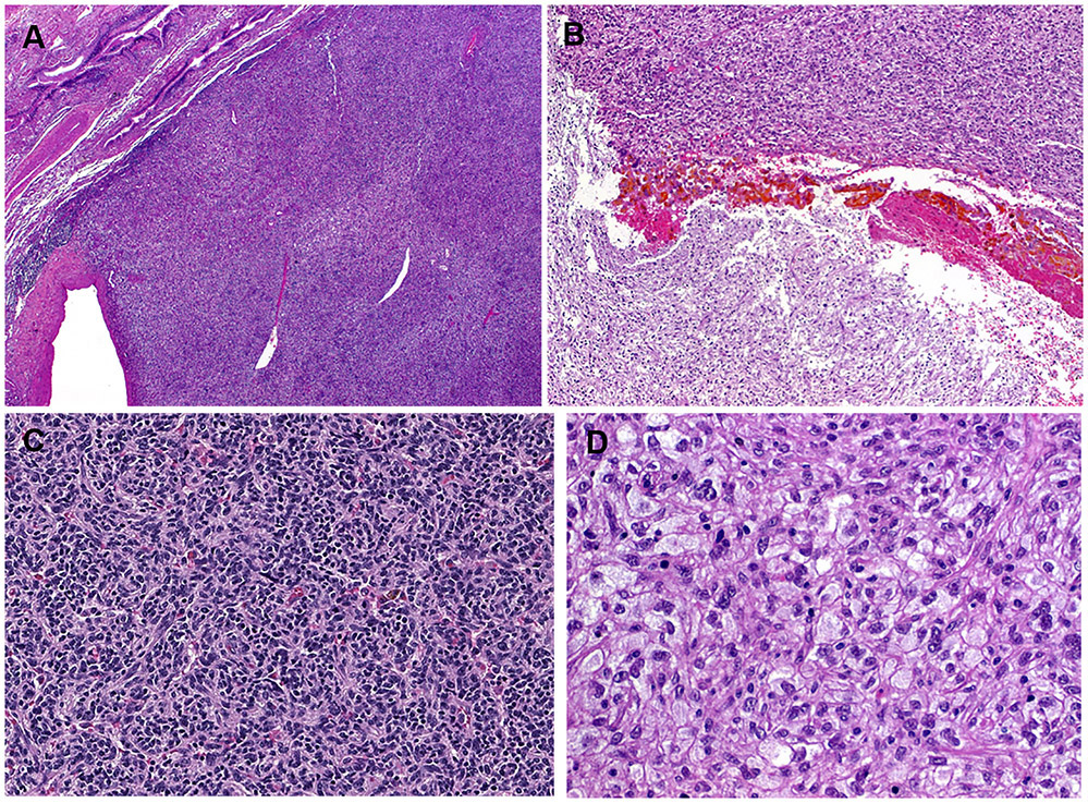
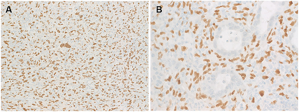
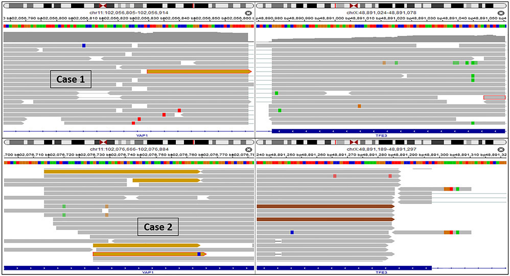
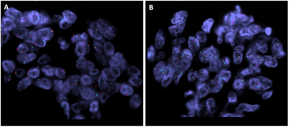
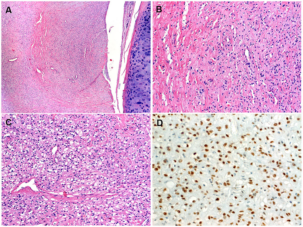
Clear cell (hemangioblastoma-like) stromal tumor of the lung (CCST-L) is a recently described distinctive rare pulmonary neoplasm of unknown histogenesis and molecular pathogenesis. Only 7 cases have been reported in 2 recent studies, although additional cases might have been reported under the heading of extraneural pulmonary hemangioblastoma. We herein describe 4 CCST-L cases, 3 of them harboring a YAP1-TFE3 fusion. The fusion-positive tumors occurred in 3 women, aged 29, 56, and 69 years. All presented with solitary lung nodules measuring 2.3 to 9.5 cm. Histologically, all tumors showed similar features being composed of relatively uniform medium-sized epithelioid to ovoid cells with clear cytoplasm and small round monomorphic nuclei. Scattered larger cells with enlarged hyperchromatic nuclei and marked pleomorphism were noted in 2 cases. The tumors were associated with a hypervascularized stroma with variable but essentially subtle resemblance to capillary hemangioblastoma and perivascular epithelioid cell tumor (PEComa). Immunohistochemistry was negative for all lineage-specific markers. Targeted RNA sequencing showed a YAP1-TFE3 fusion in 3 of 4 cases. All 3 tumors showed homogeneous nuclear TFE3 immunoreactivity. Two patients were disease free at 36 and 12 months. The third patient had biopsy-proven synchronous renal and hepatic metastases, but extended follow-up is not available (recent case). The fourth case lacking the fusion affected a 66-year-old woman and showed subtle histologic differences from the fusion-positive cases, but had comparable TFE3 immunoreactivity. CCST-L represents a distinctive entity unrelated to hemangioblastoma and likely driven by recurrent YAP1-TFE3 fusions in most cases. The relationship of our cases to the recently reported "hemangioblastoma-like" CCST-L remains to be determined. Analysis of larger series is paramount to delineate the morphologic spectrum and biological behavior of this poorly characterized entity.
Copyright © 2021 Wolters Kluwer Health, Inc. All rights reserved.
Conflict of interest statement
Conflicts of Interest and Source of Funding: Supported in part by: P50 CA217694 (C.R.A.), P50 CA140146 (C.R.A.), P30 CA008748 (C.R.A.), Cycle for Survival (C.R.A.). The authors have disclosed that they have no significant relationships with, or financial interest in, any commercial companies pertaining to this article.
Figures


References
-
- Bisceglia M, Muscarella LA, Galliani CA, Zidar N, Ben-Dor D, Pasquinelli G, la Torre A, Sparaneo A, Fanburg-Smith JC, Lamovec J, Michal M, Bacchi CE. Extraneuraxial Hemangioblastoma: Clinicopathologic Features and Review of the Literature. Adv Anat Pathol. 2018;25:197–215. - PubMed
-
- Michal M, Vanecek T, Sima R, et al. Primary capillary hemangioblastoma of peripheral soft tissues. Am J Surg Pathol. 2004;28:962–966. - PubMed
-
- Doyle LA, Fletcher CD. Peripheral hemangioblastoma: clinicopathologic characterization in a series of 22 cases. Am J Surg Pathol 2014;38:119–27. - PubMed
-
- Lindholm KE, Moran CA. Hemangioblastoma-like Clear Cell Stromal Tumor of the Lung: A Clinicopathologic and Immunohistochemical Study of 5 Cases. Am J Surg Pathol. 2020;44:771–775. - PubMed
Publication types
MeSH terms
Substances
Grants and funding
LinkOut - more resources
Full Text Sources
Other Literature Sources
Medical
Molecular Biology Databases
