

Superficial and Deep Macula Vessel Density in Healthy, Glaucoma Suspect, and Glaucoma Eyes
- PMID: 33899812
- PMCID: PMC8169636
- DOI: 10.1097/IJG.0000000000001860
Superficial and Deep Macula Vessel Density in Healthy, Glaucoma Suspect, and Glaucoma Eyes
Abstract
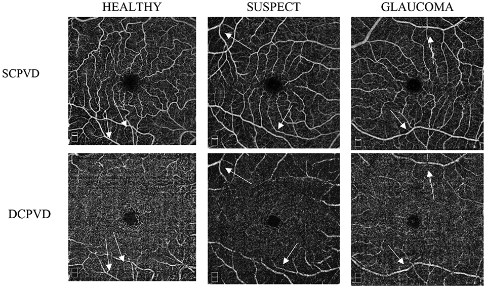
Precis: Macular superficial capillary plexus (SCP) vessel density is more informative than deep capillary plexus (DCP) vessel density for the detection of glaucoma.
Purpose: The purpose of this study was to characterize optical coherence tomography angiography macular SCP and projection-resolved DCP vessel densities and compare their diagnostic accuracies with ganglion cell complex (GCC) thickness in healthy, glaucoma suspect, and glaucoma eyes.
Materials and methods: Sixty-eight eyes of 44 healthy subjects, 26 eyes of 16 preperimetric glaucoma suspects, and 161 eyes of 124 glaucoma patients from the Diagnostics Innovations in Glaucoma Study with good quality high-density 6×6 mm2 macula optical coherence tomography angiography images were included. The diagnostic accuracy of SCP vessel density, projection-resolved DCP vessel density and GCC thickness were compared among groups.
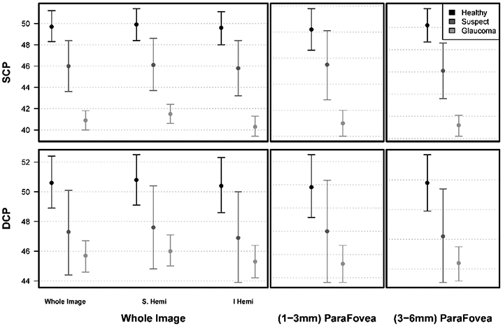
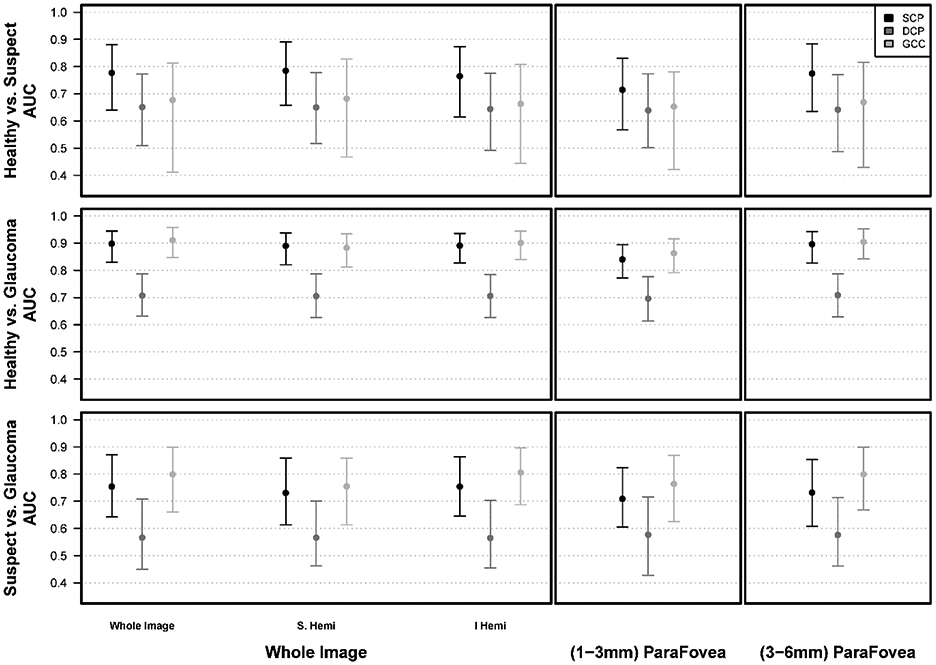
Results: Mean whole image vessel density (wiVD; % of area occupied by vessels containing flowing blood) in the SCP layer was highest in healthy eyes (49.7%), followed by glaucoma suspect eyes (46.0%), and glaucoma eyes (40.9%) (P<0.001). Mean wiVD in the DCP layer was similar in healthy (50.6%), glaucoma suspect (47.3%), and glaucoma eyes (45.7%) (P=0.925). Diagnostic accuracy of both GCC thickness and SCP wiVD was significantly higher than DCP wiVD for classifying healthy and glaucoma eyes [adjusted area under the receiver operating characteristic curve (95% confidence interval): GCC=0.86 (0.72, 0.94), SCP=0.80 (0.66, 0.91) and DCP=0.44 (0.30, 0.57)] (P<0.001).
Conclusions: SCP vessel densities have better diagnostic accuracy for detecting glaucoma than DCP vessel densities. Although the diagnostic accuracy of the macula SCP is relatively modest, it is more informative than the DCP.
Copyright © 2021 Wolters Kluwer Health, Inc. All rights reserved.
Conflict of interest statement
Disclosure: P.I.C.M. was previously employed at Heidelberg Engineering, Alcon. L.M.Z. received financial support from National Eye Institute, Carl Zeiss Meditec Inc., Heidelberg Engineering GmbH, Optovue Inc., Topcon Medical Systems Inc. T.S. was a recipient at Alcon. R.N.W. received financial support from National Eye Institute, Heidelberg Engineering, Carl Zeiss Meditec, Konan, Optovue, Topcon, Centervue; is a consultant for Aerie Pharmaceuticals, Alcon, Allergan, Bausch & Lomb, Eyenovia. The remaining authors declare no conflict of interest.
Figures

Comment in
-
Response to Letter to the Editor: Superficial and Deep Macula Vessel Density in Healthy, Glaucoma Suspect, and Glaucoma Eyes.J Glaucoma. 2021 Dec 1;30(12):1082-1083. doi: 10.1097/IJG.0000000000001951. J Glaucoma. 2021. PMID: 34628427 Free PMC article. No abstract available.
-
Letter to the Editor: Superficial and Deep Macula Vessel Density in Healthy, Glaucoma Suspect, and Glaucoma Eyes.J Glaucoma. 2021 Dec 1;30(12):1082. doi: 10.1097/IJG.0000000000001952. J Glaucoma. 2021. PMID: 34628428 No abstract available.
Similar articles
-
Optical Coherence Tomography Angiography Macular Vascular Density Measurements and the Central 10-2 Visual Field in Glaucoma.J Glaucoma. 2018 Jun;27(6):481-489. doi: 10.1097/IJG.0000000000000964. J Glaucoma. 2018. PMID: 29664832 Free PMC article.
-
Macula Vessel Density and Thickness in Early Primary Open-Angle Glaucoma.Am J Ophthalmol. 2019 Mar;199:120-132. doi: 10.1016/j.ajo.2018.11.012. Epub 2018 Nov 26. Am J Ophthalmol. 2019. PMID: 30496723 Free PMC article.
-
Optical Coherence Tomography Angiography Vessel Density in Healthy, Glaucoma Suspect, and Glaucoma Eyes.Invest Ophthalmol Vis Sci. 2016 Jul 1;57(9):OCT451-9. doi: 10.1167/iovs.15-18944. Invest Ophthalmol Vis Sci. 2016. PMID: 27409505 Free PMC article.
-
Quantitative Analysis of Microvasculature in Macular and Peripapillary Regions in Early Primary Open-Angle Glaucoma.Curr Eye Res. 2020 May;45(5):629-635. doi: 10.1080/02713683.2019.1676912. Epub 2019 Oct 14. Curr Eye Res. 2020. PMID: 31587582
-
Ganglion Cell Complex Thickness and Macular Vessel Density Loss in Primary Open-Angle Glaucoma.Ophthalmology. 2020 Aug;127(8):1043-1052. doi: 10.1016/j.ophtha.2019.12.030. Epub 2020 Jan 13. Ophthalmology. 2020. PMID: 32085875 Free PMC article.
Cited by
-
Comparison of the Effects of Latanoprostene Bunod and Timolol on Retinal Blood Vessel Density: A Randomized Clinical Trial.Am J Ophthalmol. 2022 Sep;241:120-129. doi: 10.1016/j.ajo.2022.04.022. Epub 2022 May 6. Am J Ophthalmol. 2022. PMID: 35526590 Free PMC article. Clinical Trial.
-
The characteristics of fundus microvascular alterations in the course of glaucoma: a narrative review.Ann Transl Med. 2022 May;10(9):527. doi: 10.21037/atm-21-5695. Ann Transl Med. 2022. PMID: 35928752 Free PMC article. Review.
-
Multilayer Macula Vessel Density and Visual Field Progression in Glaucoma.Am J Ophthalmol. 2022 May;237:193-203. doi: 10.1016/j.ajo.2021.11.018. Epub 2021 Nov 19. Am J Ophthalmol. 2022. PMID: 34801510 Free PMC article.
-
Association of macular OCT and OCTA parameters with visual acuity in glaucoma.Br J Ophthalmol. 2023 Nov;107(11):1652-1657. doi: 10.1136/bjo-2022-321460. Epub 2022 Sep 9. Br J Ophthalmol. 2023. PMID: 36126109 Free PMC article.
-
Relationships of Macular Functional Impairment With Structural and Vascular Changes According to Glaucoma Severity.Invest Ophthalmol Vis Sci. 2023 Sep 1;64(12):5. doi: 10.1167/iovs.64.12.5. Invest Ophthalmol Vis Sci. 2023. PMID: 37669065 Free PMC article.
References
-
- Halpern DL, Grosskreutz CL. Glaucomatous optic neuropathy: mechanisms of disease. Ophthalmol Clin North Am. 2002;15(1):61–68. - PubMed
-
- Flammer J, Orgül S, Costa VP, et al. The impact of ocular blood flow in glaucoma. Prog Retin Eye Res. 2002;21(4):359–393. - PubMed
-
- Curcio CA, Allen KA. Topography of ganglion cells in human retina. J Comp Neurol. 1990;300(1):5–25. - PubMed
Publication types
MeSH terms
Grants and funding
LinkOut - more resources
Full Text Sources
Other Literature Sources
Medical
Miscellaneous
