Review
. 2021 Aug 19;16(16):2441-2451.
doi: 10.1002/cmdc.202100201. Epub 2021 May 19.

Self-Assembled Porphyrinoids: One-Component Nanostructured Photomedicines

Affiliations
Review

Self-Assembled Porphyrinoids: One-Component Nanostructured Photomedicines

Irene Paramio et al. ChemMedChem. .

Abstract

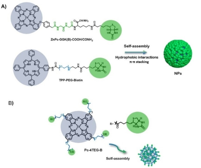
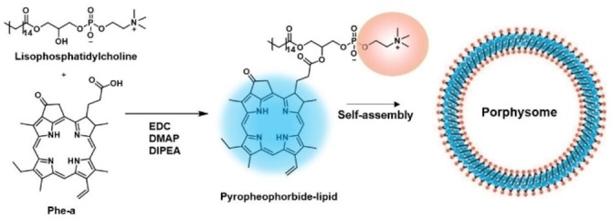
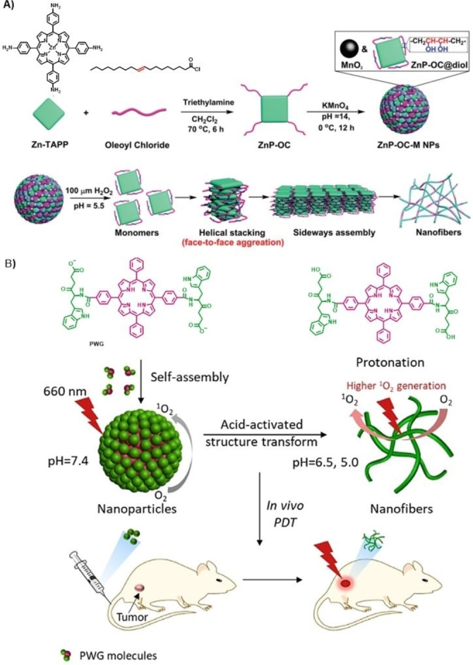
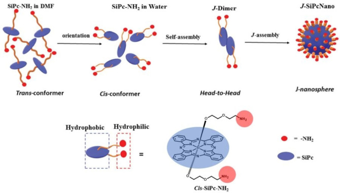
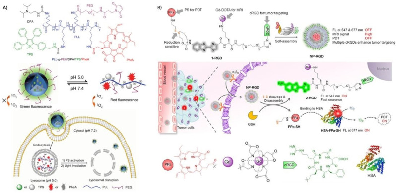
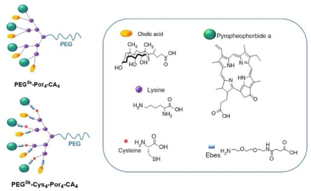
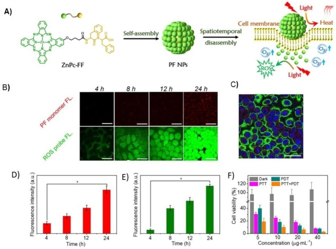
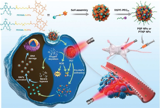
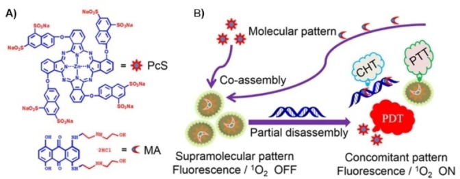
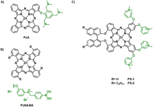
Photodynamic therapy (PDT) is becoming a promising way to treat various kinds of cancers, with few side effects. Porphyrinoids are the most relevant photosensitizers (PS) in PDT, because they present high extinction coefficients, biocompatibility, and excellent photochemical behavior. To maximize therapeutic effects, polymer-PS conjugates, and PS-loaded nanoparticles have been developed, with insights in improving tumor delivery. However, some drawbacks such as non-biodegradability, multistep fabrication, and low reagent loadings limit their clinical application. A novel strategy, noted by some authors as the "one-for-all" approach, is emerging to circumvent the use of additional delivery agents. This approach relies on the self-assembly of amphiphilic PS to fabricate nanostructures with improved transport properties. In this review we focus on different rational designs of porphyrinoid PS to achieve some of the following attributes in nanoassembly: i) selective uptake, through the incorporation of recognizable biological vectors; ii) responsiveness to stimuli; iii) combination of imaging and therapeutic functions; and iv) multimodal therapy, including photothermal or chemotherapy abilities.

Keywords: nanostructure; nanotheranostics; phototherapy; porphyrinoids; self-assembly.


Conflict of interest statement

The authors declare no conflict of interest.

Figures

Figure 1
Figure 1
Representation of the chemical structure and aggregated nanoparticles (NPs) of A) TPP‐PEG‐Biotin and ZnPc‐GGK(B)‐COOH/CONH2 , and B) Pc‐4TEG‐B.
Figure 2
Figure 2
A) Synthesis of porphysome subunit, pyropheophorbide‐lipid (left) and the schematic representation of the assembled nanovesicle (right).
Figure 3
Figure 3
A) Preparation of ZnP‐OC‐M NPs (above) and schematic illustration for the supramolecular self‐assembled nanofiber formation (below). Adapted with permission from Ref. [36]. Copyright 2020, Royal Society of Chemistry. B) Schematic illustration of self‐assembly and fibrillar transformation of the acid‐activated peptide‐porphyrin (PWG) nanoparticles and their application in PDT. Adapted with permission from Ref. [39]. Copyright 2020, Wiley‐VCH.
Figure 4
Figure 4
Schematic representation of the self‐assembly mechanism of J‐SiPcNano formation in water. Adapted with permission from Ref. [53]. Copyright 2020, Royal Society of Chemistry.
Figure 5
Figure 5
A) Chemical structure of PLL‐g‐PEG/DAP/TPS/PheA (above); Schematic illustration of the pH‐activation and its use for self‐tracking, cancer cell imaging, phototoxicity restoration in the acidic lysosome, and in situ monitoring of lysosomal membrane disruption as an indicator of therapeutic response and cell death prediction (below). Adapted with permission from Ref. [56]. Copyright 2015, Wiley‐VCH. B) Proposed self‐assembled structure of NP‐RGD (above); Schematic illustration of NP‐RGD for on demand PDT via activation by intracellular GSH and albumin (middle); Chemical structures of the PPa, DOTA‐Gd chelate, cRGD and a cartoon illustration of HSA structure (below). Adapted with permission from Ref. [41]. Copyright 2020, Wiley‐VCH.
Figure 6
Figure 6
Schematic illustration of PEG5k‐Por4‐CA4 , the cross‐linkable porphyrin‐telodendrimer PEG5k‐Cys4‐Por4‐CA4 , and the chemical structure of the building blocks that conform the telodendrimers.
Figure 7
Figure 7
A) Schematic illustration of the spatio‐temporally coupled photoactivity of PF self‐assemblies for localized adaptive tumor theranostics. B) CLSM images of MCF‐7cells incubated with PF nanoparticles (PF NPs). Red fluorescence was from monomeric Pc molecules. Green fluorescence showed the generated ROS in situ within cells using DCFH‐DA as an indicator. C) image of MCF‐7 cells incubated with PF NPs for 24 h D) Fluorescence intensity obtained from the monomeric Pc molecules within MCF‐7 cells in Figure A). E) Fluorescence intensity obtained from the ROS probe within MCF‐7 cells in Figure A). F) Viability of MCF‐7 cells incubated with PF NPs in the dark and under laser irradiation. Adapted with permission from Ref. [61]. Copyright 2019, Wiley‐VCH.
Figure 8
Figure 8
Schematic representation of the formation of heterodimeric prodrug‐nanoparticles (PSP NPs and PTKP NPs), their light‐triggered dual‐modality activation, and the PDT‐induced activation of the HAP PR104A. Adapted with permission from Ref. [62]. Copyright 2020, Royal Society of Chemistry.
Figure 9
Figure 9
A) chemical structure of octasulfonated phthalocyanine (PcS) and mitoxantrone (MA) B) Schematic illustration of the co‐assembly between PcS and MA to form a nanotheranostic agent and its nucleic‐acid‐driven activatable properties for fluorescent imaging and PDT synergized with PTT and CHT. Adapted with permission from Ref. [63]. Copyright 2018, American Chemical Society.
Figure 10
Figure 10
The chemical structure of A) PcA, B) PcN4‐BA, and C) PS‐1 and PS‐2.

References

    1. Castano A. P., Demidova T. N., Hamblin M. R., Photodiagn. Photodyn. Ther. 2004, 1, 279–293. - PMC - PubMed
    1. Castano A. P., Demidova T. N., Hamblin M. R., Photodiagn. Photodyn. Ther. 2005, 2, 1–23. - PMC - PubMed
    1. Brown S. B., Brown E. A., Walker I., Lancet Oncol. 2004, 5, 497–508. - PubMed
    1. Dai T., Huang Y. Y., Hamblin M. R., Photodiagn. Photodyn. Ther. 2009, 6, 170–188. - PMC - PubMed
    1. Rodríguez-Amigo B., Planas O., Bresolí-Obach R., Torra J., Ruiz-González R., Nonell S. in Photodynamic Medicine: From Bench to Clinic, (Eds.: Kostron H., Hasan T.), The Royal Society of Chemistry, 2016, pp. 23–62.

Publication types

MeSH terms

LinkOut - more resources