. 2021 Feb;14(s4):S57-S65.
doi: 10.14444/7166. Epub 2021 Jan 18.

Hemangioma of the Lumbar Spine Involving the Spinous Process: A Rare Case Report and Review of the Literature

Affiliations

Hemangioma of the Lumbar Spine Involving the Spinous Process: A Rare Case Report and Review of the Literature

Faisal Mohammed Saleh Konbaz et al. Int J Spine Surg. 2021 Feb.

Abstract

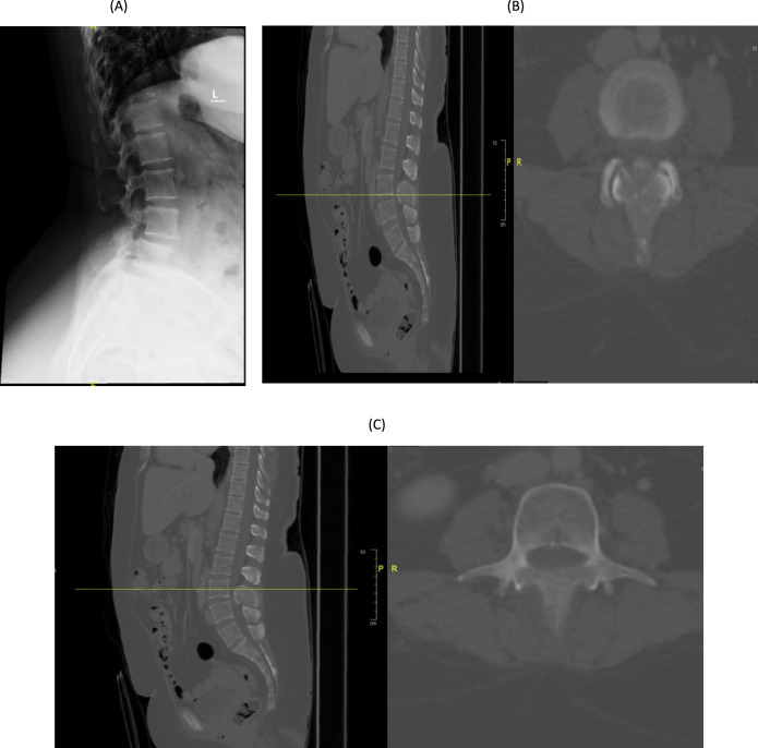
Hemangiomas of the spine are usually benign and asymptomatic. They can cause devastating complications such as pathological fractures of the spine and neurological disability. This report documents an atypical location of a hemangioma in a lumbar spinous process, in combination with a spondylolisthesis at the same level, which makes it even more uncommon. Surgery can be effective and safe and can significantly improve patient outcomes. Moreover, prior embolization can prevent acute hemorrhage in addition to providing careful diagnosis and evaluation.

Keywords: embolization; hemangioma; spinous process hemangioma; spondylolisthesis; surgery; vertebral body hemangioma.


Conflict of interest statement

Disclosures and COI: The authors received no funding for this study and report no conflicts of interest.

Figures

Figure 1
Figure 1
(A) Sagittal lumbar spine x-ray demonstrates loss of normal outline of the spinous process. (B, C) Sagittal and axial spine computed tomography (CT), respectively, demonstrating a lesion involving the lamina and spinous process of L3 vertebra.
Figure 2
Figure 2
(A) T1-weighted sagittal lumbar spine MRI with contrast showing expansile enhancing lesion involving the spinous process of L3. (B) T1-weighted postcontrast axial image of the L3 lumbar vertebra. (C) T1-weighted coronal MRI with contrast.
Figure 3
Figure 3
(A) Right L3 segmental artery angiogram anteroposterior view demonstrating abnormal vascular blush and contrast pooling consistent with hemangioma. (B) Lateral view angiogram. (C) Fluoroscopic spot image obtained during spinal angiogram demonstrated embolization material (Onyx 18) in the spinous process lesion. (D) Spine computed tomography (CT) sagittal view showing embolization material (Onyx 18) seen in the spinous process of the L3 vertebra (postembolization).
Figure 4
Figure 4
Intraoperative image after decompression and excision of the tumor.
Figure 5
Figure 5
Histological examination reveals variable sizes of cystically dilated, thin-walled blood vessels that are lined by bland endothelial cells. No marked atypia, mitosis, or necrosis seen. Diagnosis: bland vascular proliferation consistent with hemangioma.
Figure 6
Figure 6
(A) Postoperative sagittal and axial computed tomography (CT) spine at L2 pedicle. (B) Postoperative sagittal and axial CT spine at L3 pedicle. (C–E) Postoperative sagittal and axial CT spine at L4 pedicle. Axial image showing the amount of tumor that was resected including the medial wall of the left L3 pedicle.
Figure 6
Figure 6
Continued.
Figure 7
Figure 7
(A, B) Anteroposterior and lateral postoperative x-rays, respectively, showing 2 rods and transpedicular screws seen transfixing the vertebral body from L2 to L4. No evidence of looseness or alignment deformity. There is no pedicle screw on the left L3 vertebra.
Figure 8
Figure 8
(A, B) Anteroposterior and lateral views of follow-up x-rays, respectively, 2 years postoperatively showing 22 rods and transpedicular screws seen transfixing the vertebral body from L2 to L4. No evidence of looseness or alignment deformity. There is no pedicle screw on the left L3 vertebra.

References

    1. Doppman JL, Oldfield EH, Heiss JD. Symptomatic vertebral hemangiomas: treatment by means of direct intralesional injection of ethanol. Radiology. doi: 10.1148/radiology.214.2.r00fe46341. - DOI - PubMed
    1. Peckham ME, Hutchins TA. Imaging of vascular disorders of the spine. Radiol Clin. 2019;57(2):307–318. doi: 10.1016/j.rcl.2018.09.005. - DOI - PubMed
    1. Theodorou DJ, Theodorou SJ, Sartoris DJ. An imaging overview of primary tumors of the spine: part 1. Benign tumors. Clin Imaging. doi: 10.1016/j.clinimag.2007.09.011. - DOI - PubMed
    1. Yochum TR, Lile RL, Schultz GD, Mick TJ, Brown CW. Acquired spinal stenosis secondary to an expanding thoracic vertebral hemangioma. Spine (Phila Pa 1976) doi: 10.1097/00007632-199302000-00021. - DOI - PubMed
    1. Malawski S, Sokólski B. A case of spinal hemangioma uncommonly located within spinous processes and laminae of Th12. Chir Narzadow Ruchu Ortop Pol. 1996;61(1):47–51. - PubMed

LinkOut - more resources