. 2021 Apr 26;13(9):12308-12333.
doi: 10.18632/aging.203010. Epub 2021 Apr 26.

Shikimic acid protects skin cells from UV-induced senescence through activation of the NAD+-dependent deacetylase SIRT1

Affiliations

Shikimic acid protects skin cells from UV-induced senescence through activation of the NAD+-dependent deacetylase SIRT1

Alfredo Martínez-Gutiérrez et al. Aging (Albany NY). .

Abstract

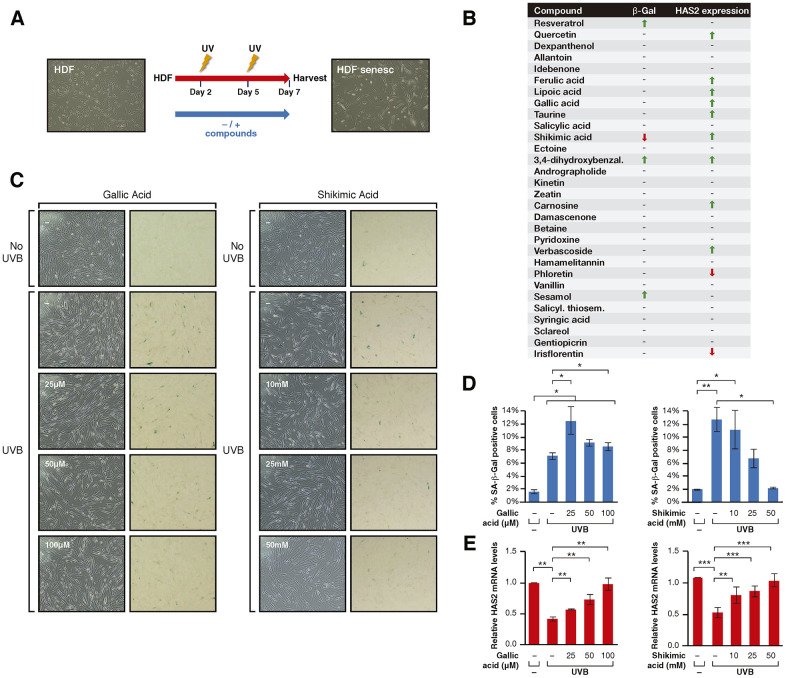
UV radiation is one of the main contributors to skin photoaging by promoting the accumulation of cellular senescence, which in turn induces a proinflammatory and tissue-degrading state that favors skin aging. The members of the sirtuin family of NAD+-dependent enzymes play an anti-senescence role and their activation suggests a promising approach for preventing UV-induced senescence in the treatment of skin aging. A two-step screening designed to identify compounds able to protect cells from UV-induced senescence through sirtuin activation identified shikimic acid (SA), a metabolic intermediate in many organisms, as a bona-fide candidate. The protective effects of SA against senescence were dependent on specific activation of SIRT1 as the effect was abrogated by the SIRT1 inhibitor EX-527. Upon UV irradiation SA induced S-phase accumulation and a decrease in p16INK4A expression but did not protect against DNA damage or increased polyploidies. In contrast, SA reverted misfolded protein accumulation upon senescence, an effect that was abrogated by EX-527. Consistently, SA induced an increase in the levels of the chaperone BiP, resulting in a downregulation of unfolded protein response (UPR) signaling and UPR-dependent autophagy, avoiding their abnormal hyperactivation during senescence. SA did not directly activate SIRT1 in vitro, suggesting that SIRT1 is a downstream effector of SA signaling specifically in the response to cellular senescence. Our study not only uncovers a shikimic acid/SIRT1 signaling pathway that prevents cellular senescence, but also reinforces the role of sirtuins as key regulators of cell proteostasis.

Keywords: SIRT1; UV irradiation; human dermal fibroblasts; senescence; shikimic acid.


Conflict of interest statement

CONFLICTS OF INTEREST: A.M and J.M are employees of mesoestetic Pharma Group, s.l. Mesoestetic Pharma Group, s.l has applied for a patent, PCT 2019/070704, for the use of shikimic acid in the treatment of skin aging.

Figures

Figure 1
Figure 1
(A) Schematic representation of the compound testing for sirtuin activity evaluation in HDF. Cells were treated with or without the compounds for 24h and harvested for analysis. (B) Sirtuin activity assay results including H4K16 and H3K9 acetylation for cells treated with each compound. The results are shown by arrows that indicate increase/decrease (↑, ↓) and hyphens that indicate no effect (-). (C) Western Blot analysis and (D) quantification of H4K16ac and H3K9ac levels in cells treated with Resveratrol or Shikimic acid at the indicated concentrations. H4K16 and H3K9 levels were normalized to H4 and H3 levels respectively. Student T-test, *p<0.05, **p<0.01, ***p<0.001.
Figure 2
Figure 2
(A) Schematic representation of the protocol for senescence induction using UVB irradiation. Cells were treated -/+ the compounds from day 1 to 7 after seeding, irradiated with UVB on days 2 and 5 and harvested for analysis on day 7. (B) Senescence assay results including β-Gal staining and HAS2 gene expression analysis for cells treated with each compound. The results are shown by arrows that indicate increase/decrease (↑, ↓) and hyphens that indicate no effect (-). (C) Phase-contrast (left column) and bright field (right column) microscope images of SA-β-Gal staining showing non-irradiated cells, UVB-irradiated cells non-treated and treated with Gallic acid or Shikimic acid at the indicated concentrations. (D) Percentage of SA-β-Gal positive cells and (E) Relative HAS2 mRNA levels in non-irradiated cells, UVB-irradiated cells and UVB-irradiated cells treated with Gallic acid or Shikimic acid at the indicated concentrations. Student T-test, *p<0.05, **p<0.01, ***p<0.001.
Figure 3
Figure 3
(A) Schematic representation of the protocol for senescence induction using UVB irradiation and study of EX527 involvement in the effect of Shikimic acid. Cells were treated with or without shikimic acid -/+ EX-527 from day 1 until day 7 after seeding, irradiated with UVB on days 2 and 5 and harvested for analysis on day 7. (B) Phase-contrast (left column) and bright field (right column) microscope images of β-Gal staining showing non-irradiated cells non-treated and treated with shikimic acid (10, 25 and 50 mM), UVB-irradiated cells non-treated and treated with shikimic acid (10, 25 and 50 mM), shikimic acid (10, 25 and 50 mM) + EX-527 1 μM and EX-527 1 μM. (C) Percentage of SA-β-Gal positive cells and (D) relative HAS2 mRNA levels in non-irradiated cells non-treated and treated with Shikimic acid (10, 25 and 50 mM), UVB-irradiated cells non-treated and treated with Shikimic acid (10, 25 and 50 mM), Shikimic acid (10, 25 and 50 mM) + EX-527 1 μM and EX-527 1 μM. (E) Percentage of SA-β-Gal as in (C) of SA 25 or 50mM upon siRNA mediated downregulation of SIRT1, SIRT2 or SIRT6. siScr: scramble siRNA. Student T-test, *p<0.05, **p<0.01, ***p<0.001. (F) Levels of H3K9ac/H3 and H4K16ac/H4 under UV-induced senescence in HFD incubated with -/+25mM SA -/+1μM EX-527. (G) IL-6 mRNA levels of the assays as in (D). (H) The activity of FLAG-SIRT1 purified from HEK293 cells was tested in in vitro deacetylation assays -/+ NAD+ -/+ Shikimic acid (1 μM-10mM) in presence of hyperacetylated core histones. SIRT1 activity was then evaluated by analyzing acetylation of H3K9, H4K16 and p53K382 by Western blot. Student T-test, *p<0.05, **p<0.01, ***p<0.001.
Figure 4
Figure 4
(A) SRB and Trypan blue cell viability assays performed in cells treated with or without Shikimic acid (5, 10, 25 and 50 mM) for 24h. (B) Apoptosis detection using the Annexin V-FITC/PI double staining followed by flow cytometry in non-irradiated and non-treated cells, irradiated and non-treated cells and irradiated cells treated with Shikimic acid (10, 25 and 50 mM). (C) Representation and statistical analysis of the percentage of apoptotic cells in each of the conditions analyzed. (D) Cell cycle analysis using PI staining followed by flow cytometry. (E) Relative p16INK4a mRNA levels monitored by qPCR in the indicated conditions. Student T-test, *p<0.05. (F) Immunofluorescence of γH2AX in non-irradiated and non-treated cells, irradiated and non-treated cells and irradiated cells treated with Shikimic acid (10, 25 and 50 mM). (G) Principal component analysis (PCA) on Savitzky–Golay second derivatized spectra in the fingerprint region (1800–950 cm−1) for non-irradiated cells (No UVB), irradiated non-treated cells (UVB), irradiated cells treated with Shikimic acid 25 mM (UVB SA) and irradiated cells treated with Shikimic acid 25 mM plus EX-527 1 μM (UVB SA+EX-527). Student T-test, *p<0.05, **p<0.01, ***p<0.001. (H) Beta-sheet to alpha-helix ratio obtained by curve-fitting analysis of the amide I band (1700-1600 cm-1).
Figure 5
Figure 5
(A) Western-blot of BiP proteins levels in UVB or non UVB irradiated cells treated with SA at the indicated concentrations in presence or absence of EX-527 1 μM. (B) Quantification of n=3 experiments as (A). The levels of BiP are normalized by the loading control (histone H3) and represented related to the conditions in UVB irradiated cells in absence of SA and EX-527. Student T-test, *p<0.05. (C) Percentage of SA-β-Gal in assays performed as in Figure 3C. (D) Levels of spliced and unspliced XBP1 mRNA monitored by qPCR analysis in cells treated in the indicated conditions in presence or absence of SA and/or EX-527. The levels were normalized by internal controls and represented in each case related to the conditions in UVB irradiated cells in absence of SA and EX-527. Student T-test, *p<0.05, **p<0.01, ***p<0.001. (E) LC3-II protein levels in experiments like in (A). The levels of LC3-II are normalized by the loading control (tubulin) and represented related to the conditions in UVB irradiated cells in absence of SA and EX-527. Student T-test, *p<0.05. (F) Proposed Model for the effect of SA on senescence through SIRT1. Other possible unexplored connections between SA and senescence are indicated by blue broken lines.

References

    1. Sjerobabski-Masnec I, Situm M. Skin aging. Acta Clin Croat. 2010; 49:515–18. - PubMed
    1. Kohl E, Steinbauer J, Landthaler M, Szeimies RM. Skin ageing. J Eur Acad Dermatol Venereol. 2011; 25:873–84. 10.1111/j.1468-3083.2010.03963.x - DOI - PubMed
    1. Ganceviciene R, Liakou AI, Theodoridis A, Makrantonaki E, Zouboulis CC. Skin anti-aging strategies. Dermatoendocrinol. 2012; 4:308–19. 10.4161/derm.22804 - DOI - PMC - PubMed
    1. Uitto J. Understanding premature skin aging. N Engl J Med. 1997; 337:1463–65. 10.1056/NEJM199711133372011 - DOI - PubMed
    1. Pandel R, Poljšak B, Godic A, Dahmane R. Skin photoaging and the role of antioxidants in its prevention. ISRN Dermatol. 2013; 2013:930164. 10.1155/2013/930164 - DOI - PMC - PubMed

Publication types