. 2021 Apr 26;16(4):e0250822.
doi: 10.1371/journal.pone.0250822. eCollection 2021.

Beyond dissolution: Xerostomia rinses affect composition and structure of biomimetic dental mineral in vitro

Affiliations

Beyond dissolution: Xerostomia rinses affect composition and structure of biomimetic dental mineral in vitro

Mikayla M Moynahan et al. PLoS One. .

Abstract

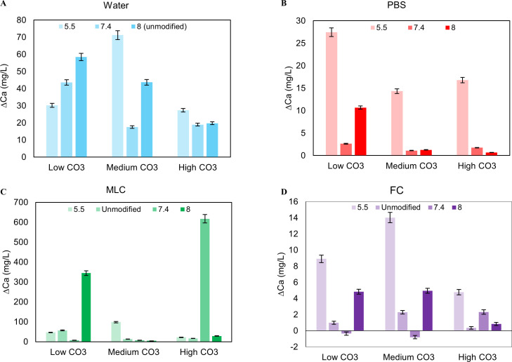
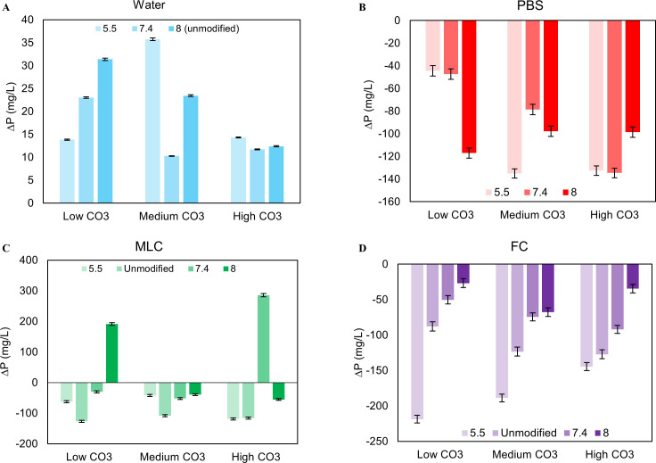
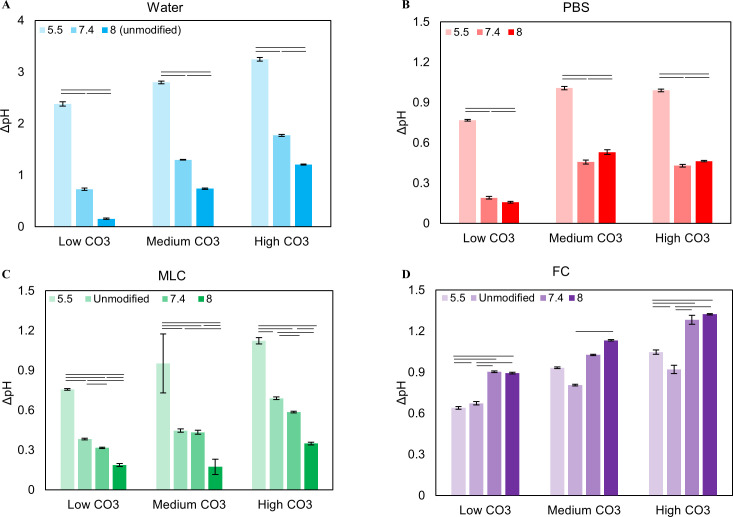
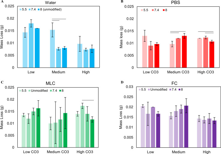
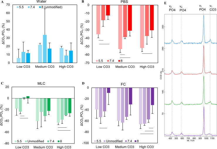
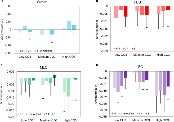
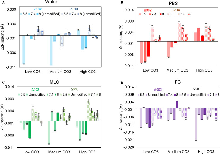
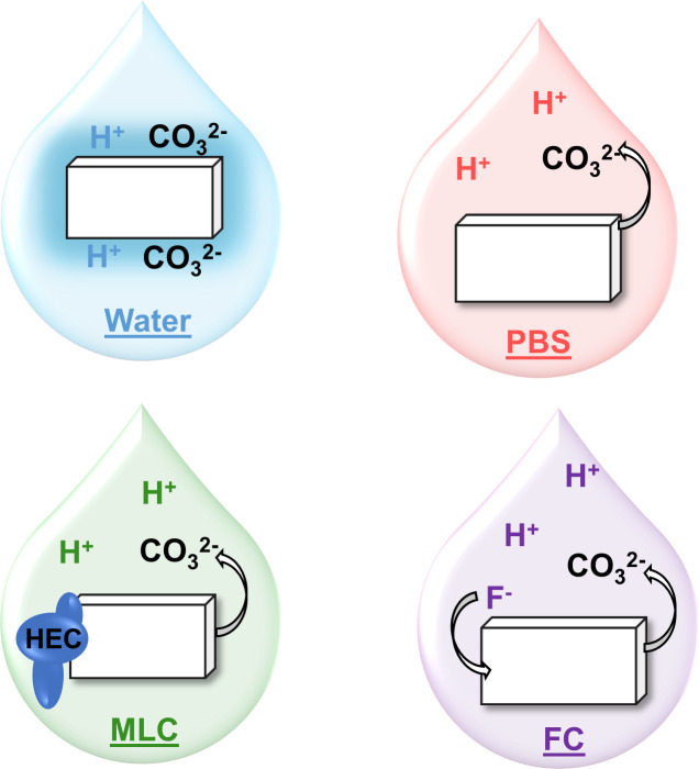
Xerostomia, known as dry mouth, is caused by decreased salivary flow. Treatment with lubricating oral rinses provides temporary relief of dry mouth discomfort; however, it remains unclear how their composition affects mineralized dental tissues. Therefore, the objective of this study was to analyze the effects of common components in xerostomia oral rinses on biomimetic apatite with varying carbonate contents. Carbonated apatite was synthesized and exposed to one of the following solutions for 72 hours at varying pHs: water-based, phosphorus-containing (PBS), mucin-like containing (MLC), or fluoride-containing (FC) solutions. Post-exposure results indicated that apatite mass decreased irrespective of pH and solution composition, while solution buffering was pH dependent. Raman and X-ray diffraction analysis showed that the addition of phosphorus, mucin-like molecules, and fluoride in solution decreases mineral carbonate levels and changed the lattice spacing and crystallinity of bioapatite, indicative of dissolution/recrystallization processes. The mineral recrystallized into a less-carbonated apatite in the PBS and MLC solutions, and into fluorapatite in FC. Tap water did not affect the apatite lattice structure suggesting formation of a labile carbonate surface layer on apatite. These results reveal that solution composition can have varied and complex effects on dental mineral beyond dissolution, which can have long term consequences on mineral solubility and mechanics. Therefore, clinicians should consider these factors when advising treatments for xerostomia patients.


Conflict of interest statement

The authors have declared that no competing interests exist.

Figures

Fig 1
Fig 1. Change in calcium levels in the solutions after exposure.
Calcium increased in all solutions, confirming apatite dissolution (A-D). Generally, calcium levels in the solution increased as pHi decreased for PBS (B). For water, MLC, and FC, there are no apparent trends (A, C, D). Overall, water had the most calcium in the solution after exposure since there is less calcium in water compared to the other solutions (A).
Fig 2
Fig 2. Change in phosphorus amounts in the solution after exposure.
P decreased in all solutions except water, indicating an uptake of phosphorus in the apatite during recrystallization (A-D). There are no apparent trends in PBS and MLC (B, C). In FC, P decreased in the solution as initial pHi decreased (D). P increased in water due to the non-detectable levels of the initial solution (A).
Fig 3
Fig 3. The change in pH (ΔpH) of the solutions as a function of initial pH.
ΔpH increased as initial pH decreased for Water, PBS, and MLC, indicating that the powder is buffering the solution more at lower pHs (A, B, C). For FC, ΔpH increased as initial pH increased (D).
Fig 4
Fig 4. Mass loss as a function of pH at each wt% carbonate for each solution.
Mass decreased for all solutions, indicating powder erosion (A-D). Powders in FC had the greatest mass loss (D) while PBS and Water had the least (A, B).
Fig 5
Fig 5. Δ Carbonate: Phosphate ratios of CAP powders after exposure as a function of initial pH.
CO32-:PO42- ratios decreased for PBS, MLC, and FC after solution exposure (B-D) while Water increased, suggesting an increase in carbonate amount (A). For PBS, CO32-:PO42- significantly decreases as pHi decreases at all wt% CO32- (B). MLC and FC trended similarly to PBS (C, D), suggesting a loss of carbonate in apatite. Raman spectra of 6 wt% carbonated apatite after exposure in water (blue), PBS (red), MLC (green), and FC (purple) solutions at pH 5.5 (E).
Fig 6
Fig 6. The internal microstrain of CAP after solution exposure.
Small variations in microstrain were shown in water, suggesting no major change in structure (A). The microstrain decreased for PBS, MLC, and FC, although there are no trends relative to pH and wt% CO32- (B-D). This shows that the apatite is becoming a more perfect crystal after solution exposure. Error bars account for the standard deviations related to the y-intercept derived from the Halder-Wagner equation.
Fig 7
Fig 7. D-spacing of the c-axis (002) and a-axis (004) of CAP after exposure.
Generally, the c-axis decreased, and the a-axis increased for PBS and MLC (B, C). For FC, both c-axis and a-axis decreased (C). These changes in d-spacing specify structural changes in apatite after solution exposure. Water exhibited variations of these axes, which is indicative of no structural differences of CAP (A). Error bars account for XRD peak fitting errors.
Fig 8
Fig 8. Summary of dissolution mechanism of CAP in each solution.
Water does not affect apatite erosion and leaves labile carbonate on the crystal surface. The addition of phosphorus in solution allows carbonate to be released from apatite to buffer the acid in the solution. MLC has a similar mechanism, however, mucin-like molecules binding to apatite may affect apatite erosion. While carbonate is dissolved from apatite into FC to buffer the acid, fluoride may be incorporated into apatite during recrystallization.

Similar articles

Cited by

References

    1. Bartels C. Xerostomia. Available from: https://oralcancerfoundation.org/complications/xerostomia/, 2019.
    1. Sreebny LM, Vissink A. Dry Mouth: The Malevolent Symptom: A Clinical Guide. Ames, Iowa, Ames, Iowa. Wiley-Blackwell, 2010.
    1. Delli K, Spijkervet FKL, Kroese FGM, Bootsma H, Vissink A. Xerostomia. Saliva: Secretion and Functions 2014;24:109–125 10.1159/000358792 - DOI - PubMed
    1. Department of Scientific Information, ADA Science Institute. Xerostomia (Dry Mouth). Available from: https://www.ada.org/en/member-center/oral-health-topics/xerostomia, 2020.
    1. Ship JA, Pillemer SR, Baum BJ. Xerostomia and the Geriatric Patient. Journal of the American Geriatrics Society 2002;50:535–543 10.1046/j.1532-5415.2002.50123.x - DOI - PubMed

Publication types

MeSH terms