Review
. 2021 Nov:85:100969.
doi: 10.1016/j.preteyeres.2021.100969. Epub 2021 Apr 23.

The retinal pigment epithelium: Development, injury responses, and regenerative potential in mammalian and non-mammalian systems

Affiliations
Review

The retinal pigment epithelium: Development, injury responses, and regenerative potential in mammalian and non-mammalian systems

Stephanie M George et al. Prog Retin Eye Res. 2021 Nov.

Abstract

Diseases that result in retinal pigment epithelium (RPE) degeneration, such as age-related macular degeneration (AMD), are among the leading causes of blindness worldwide. Atrophic (dry) AMD is the most prevalent form of AMD and there are currently no effective therapies to prevent RPE cell death or restore RPE cells lost from AMD. An intriguing approach to treat AMD and other RPE degenerative diseases is to develop therapies focused on stimulating endogenous RPE regeneration. For this to become feasible, a deeper understanding of the mechanisms underlying RPE development, injury responses and regenerative potential is needed. In mammals, RPE regeneration is extremely limited; small lesions can be repaired by the expansion of adjacent RPE cells, but large lesions cannot be repaired as remaining RPE cells are unable to functionally replace lost RPE tissue. In some injury paradigms, RPE cells proliferate but do not regenerate a morphologically normal monolayer, while in others, proliferation is pathogenic and results in further disruption to the retina. This is in contrast to non-mammalian vertebrates, which possess tremendous RPE regenerative potential. Here, we discuss what is known about RPE formation during development in mammalian and non-mammalian vertebrates, we detail the processes by which RPE cells respond to injury, and we describe examples of RPE-to-retina and RPE-to-RPE regeneration in non-mammalian vertebrates. Finally, we outline barriers to RPE-dependent regeneration in mammals that could potentially be overcome to stimulate a regenerative response from the RPE.

Keywords: Age-related macular degeneration (AMD); Development; Regeneration; Retinal pigment epithelium (RPE); Zebrafish.


Conflict of interest statement

L.L.L. is co-inventor on a US Patent (#9,458,428) related to deriving RPE from pluripotent stem cells in vitro; while not directly related to the content herein, we wish to disclose this patent. All other authors declare no conflicts of interest.

Figures

Figure 1.
Figure 1.. RPE biology and generalized functions.
Cartoon illustrating the mature, polarized RPE monolayer and its interactions with the rod and cone PRs, BM, and CC, as well as the diversity of functions mediated by the RPE. Abbreviations: PRs, photoreceptors; RPE, retinal pigment epithelium; CC, choriocapillaris; BM, Bruch’s membrane; POS, photoreceptor outer segments.
Figure 2.
Figure 2.. Eye morphogenesis in amniotes and teleosts.
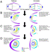
(A) In teleosts (e.g. zebrafish and medaka) and amniotes (e.g. mammals, birds), the OVs evaginate from the presumptive eye field in the forebrain. Enlargement of the left OV (black dashed boxes, A) to highlight subsequent steps of eye development (B-D). (B) OV elongation occurs posteriorly in teleosts, where RPE progenitors occupy a small portion of the medial (dorsal) layer as a pseudostratified epithelium. In amniotes, the OV extends outward, and the future RPE cells are cuboidal and occupy the region surrounding the presumptive retina. (C) Invagination of the surface ectoderm and distal OV occurs simultaneously; RPE in both teleosts and amniotes are cuboidal epithelial cells. (D) Eye morphogenesis concludes with the formation of a bilayered optic cup: RPE cells in teleosts are squamous and directly surround/interdigitate with the retina with no intervening space, while RPE cells in amniotes remain cuboidal and interdigitate apically with the retina, but remain separated from it by the intraretinal space, which disappears before birth. Abbreviations: OV, optic vesicle; RPE; retinal pigment epithelium.
Figure 3.
Figure 3.. RPE-dependent repair and regeneration in mammalian and non-mammalian systems.
Models of repair and regenerative responses in mammals (A); the newt, Cynops pyrrhogaster (B); the frog, Xenopus laevis (C); and zebrafish (D). (A) In mammals, after transgenic ablation or pharmacological injury of the RPE, remaining RPE cells are unable to regenerate. (B) In adult C. pyrrhogaster, upon retinectomy RPE cells undergo reprogramming, re-enter the cell cycle, and convert into a multipotent state through the regulation of MEKERK/β-catenin and Pax6 signaling after retinectomy. The multipotent cells segregate into two layers; the outer layer renews the RPE while the inner layer regenerates NR. (C) In adult X. laevis, upon retinectomy, with RVM present, RPE cells detach from each other and BM, express Pax6, and migrate towards the RVM to regenerate NR. RPE cells that remain attached to BM replenish the RPE layer. (D) In zebrafish, rpe65a:nfsB-eGFP-mediated transgenic ablation of large swathes of RPE results in the proliferation of injury-adjacent RPE cells that subsequently regenerate lost RPE tissue in a peripheral to central fashion. Abbreviations: NR, neural retina; BM, Bruch’s Membrane; RVM, retinal vascular membrane; RPE, retinal pigment epithelium; pRPE, peripheral retinal pigment epithelium; dRPE: dedifferentiated retinal pigment epithelium; TZ: transition zone. (B, modified from Chiba, 2014; D, modified from Hanovice et al., 2019).
Figure 4.
Figure 4.. RPE ablation paradigm in zebrafish:
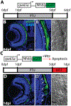
(A) Cartoon depicting the rpe65a:nfsB-eGFP transgene and treatment course of unablated embryos. (B) Transverse cryosections of an unablated 6dpf larva. (B,B’) After exposure to PTU between 1–5dpf, transgene expression is specifically restricted to mature RPE cells, with the brightest expression confined to the central two-thirds of the RPE. Arrowheads indicate apical microvilli. (B”) DIC images reveal RPE repigmentation and normal photoreceptor layer architecture. (C) Cartoon depicting the nitroreductase-mediated ablation paradigm: after washing out PTU, larvae were treated with MTZ for 24 hours. Within cells expressing the transgene, nfsB converts MTZ into a potent DNA crosslinking agent and induces cell death. (D,D’) Transverse cryosections of a 1dpi larva reveal significant disruption of eGFP+ cell morphology and disorganization in INL nuclear lamination. Arrows indicate delaminated and pyknotic nuclei. (D”) DIC images reveal a lack of RPE pigmentation and the marked disruption of photoreceptor layer architecture. Abbreviations: dpf, days post-fertilization; PTU, phenylthiourea; RPE, retinal pigment epithelium; MTZ, metronidazole; nfsB, nitroreductase; dpi, days post-injury; INL, inner nuclear layer. Green=eGFP, blue=nuclei. Dorsal is up and distal is left. Scale bar = 40mm. Figure taken from Hanovice et al., 2019.
Figure 5.
Figure 5.. Ablation of the RPE in zebrafish leads to degeneration of underlying photoreceptors.
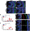
(A-D) Transverse cryosections stained for TUNEL (red). Compared to untreated (A,C) larvae, ablated RPE were disrupted by 12hpi (B), and TUNEL+ cells appeared throughout the RPE and ONL at 24hpi (D). (E, F) Quantification of TUNEL+ cells/section in the RPE (E) and ONL (F) revealed a significant increase in the RPE by 12hpi and in the ONL by 18hpi. Significance determined using Mann-Whitney U test. * p≤0.05, ** p<0.005, *** p<0.0005. (G-I) Transverse sections of unablated 6dpf larvae stained for ZPR2, an RPE marker (red, G). By 1dpi, ZPR2 is disrupted in a similar manner to eGFP (H). By 3dpi, ZPR2 signal is absent from the central injury site (I). Abbreviations: RPE, retinal pigment epithelium; hpi, hours post-injury; dpf, days post-fertilization; ONL, outer nuclear layer; TUNEL, terminal deoxynucleotidyl transferase dUTP nick end labeling. Green=eGFP, blue=nuclei. Dorsal is up and distal is left. Scale bar = 40mm. Figure modified from Hanovice et al., 2019.
Figure 6.
Figure 6.. RPE regeneration in zebrafish initiates in the periphery and proceeds inward.
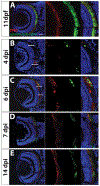
Transverse sections of unablated larvae stained for the RPE marker ZPR2 (A) at 11dpf. Ablated eyes stained for ZPR2 at 4, 6, 7 and 14dpi (B-E). Green=eGFP, blue=nuclei, red=ZPR2. eGFP+ RPE appears in the periphery at 4dpi (marked by lines at dorsal and ventral margins in B,C). As regeneration proceeds, eGFP+ RPE extends further toward the eye center, and the leading tip of the regenerated monolayer often consists of both immature and mature RPE (ZPR2+/eGFP cells in C). By 7dpi, ZPR2+ RPE is present throughout the RPE (D). By 14dpi, mature eGFP+/ZPR2+ RPE cells are present throughout the RPE (E). Dorsal is up and distal is left. Abbreviations: RPE, retinal pigment epithelium; dpf, days post-fertilization; dpi, days post-injury. Scale bar = 40mm. Figure modified from Hanovice et al., 2019.
Figure 7.
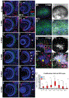
Figure 7.. RPE regeneration in zebrafish involves a robust proliferative response.
(A-L) Transverse retinal sections of unablated (A-F) and ablated (G-L) larvae exposed to 24-hour BrdU pulses at various days post-injury. BrdU+ cells first appear in the periphery as early as 0–1dpi (arrow, G), and 1–2dpi (arrow, H). As regeneration proceeds, BrdU+ cells appear closer to the central injury site and at the inner tip of the regenerating RPE layer (arrows, I). BrdU+ cells then populate the injury site (arrows, J-L). (M-T) en face wholemount images of unablated (M-P) and ablated (Q-T) eyes from larvae exposed to BrdU between 3–4dpi. White arrowheads in (Q) and (inset, Q) indicate BrdU+/eGFP+ cells near the injury site. Yellow arrowhead in (S) and (T) indicate BrdU+ cells proximal to the injury site that are beginning to become pigmented. (Inset, T) Magnified image of BrdU+, pigmented cells. (U) Quantification of total number of BrdU+ cells/section in the RPE reveals an increase of BrdU+ cells in the RPE starting at 0–1dpi and peaking at 3–4dpi. Mann-Whitney U Test, * p<0.05, ** p<0.005, *** p<0.0005. Dorsal is up and distal is left. Abbreviations: dpi, days post-injury; RPE, retinal pigment epithelium; BrdU, bromodeoxyuridine. Scale bar = 40mm. Figure modified from Hanovice et al., 2019.
Figure 8.
Figure 8.. Pharmacological inhibition of Wnt pathway activity using IWR-1 impairs RPE regeneration in zebrafish.
(A-D) Transverse sections of lef1, a marker of Wnt pathway activity, or sense RNA expression in unablated 6dpf (MTZ−) and ablated 1dpi (MTZ+) larvae. lef1 is detected in and around the RPE in MTZ+ (B’) but not MTZ- larvae (A’). lef1: n>5; lef1 sense: n=4. (E-J) Transverse sections of 4dpi ablated DMSO- (E,H; n=10) and 15μM IWR-1-treated (F,I; n=11) larvae exposed to a 24-hour pulse of BrdU from 3–4dpi. (E,F) Green=eGFP, blue=DNA, red=BrdU; white arrowheads highlight BrdU+ cells in the RPE. (G) Quantification of BrdU+ cells/section reveals that IWR-1 treatment significantly decreases the number of proliferative cells in the RPE at 4dpi (Student’s unpaired t-test, *** p<0.0001). Brightfield images (H,I) and quantification of percent RPE recovery/section (J) shows a significant delay in recovery of a pigmented monolayer in IWR-1 treated larvae (Student’s unpaired t-test, *** p<0.0001). (I) Black arrowheads indicate the central-most edge of the regenerating RPE. Abbreviations: dpf, days post-fertilization; MTZ, metronidazole; dpi, days post-injury; RPE, retinal pigment epithelium; BrdU, bromodeoxyuridine; DMSO, dimethylsulfoxide. Dorsal is up and distal is left. Scale bars = 40μm. Figure modified from Hanovice et al., 2019.

References

    1. Ail D, Perron M, 2017. Retinal Degeneration and Regeneration-Lessons From Fishes and Amphibians. Curr Pathobiol Rep 5, 67–78. - PMC - PubMed
    1. Aisenbrey S, Zhang M, Bacher D, Yee J, Brunken WJ, Hunter DD, 2006. Retinal Pigment Epithelial Cells Synthesize Laminins, Including Laminin 5, and Adhere to Them through α3- and α6-Containing Integrins. Investigative Ophthalmology & Visual Science 47, 5537–5544. - PMC - PubMed
    1. Akhtar-Schäfer I, Wang L, Krohne TU, Xu H, Langmann T, 2018. Modulation of three key innate immune pathways for the most common retinal degenerative diseases. EMBO Mol Med 10. - PMC - PubMed
    1. Al-Hussaini H, Kam JH, Vugler A, Semo M, Jeffery G, 2008. Mature retinal pigment epithelium cells are retained in the cell cycle and proliferate in vivo. Mol Vis 14, 1784–1791. - PMC - PubMed
    1. Al-Hussaini H, Kilarkaje N, Shahabi G, Al-Mulla F, 2016. Proliferation and Migration of Peripheral Retinal Pigment Epithelial Cells Are Associated with the Upregulation of Wingless-Related Integration and Bone Morphogenetic Protein Signaling in Dark Agouti Rats. Med Princ Pract 25, 408–416. - PMC - PubMed

Publication types