. 2021 Jul;34(7):e4531.
doi: 10.1002/nbm.4531. Epub 2021 Apr 26.

Free-breathing abdominal T1 mapping using an optimized MR fingerprinting sequence

Affiliations

Free-breathing abdominal T1 mapping using an optimized MR fingerprinting sequence

Max H C van Riel et al. NMR Biomed. 2021 Jul.

Abstract

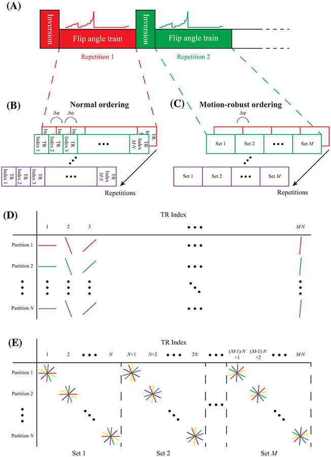
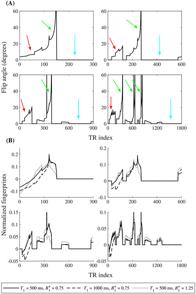
In this work, we propose a free-breathing magnetic resonance fingerprinting (MRF) method that can be used to obtain B1+ -robust quantitative T1 maps of the abdomen in a clinically acceptable time. A three-dimensional MRF sequence with a radial stack-of-stars trajectory was implemented, and its k-space acquisition ordering was adjusted to improve motion-robustness in the context of MRF. The flip angle pattern was optimized using the Cramér-Rao Lower Bound, and the encoding efficiency of sequences with 300, 600, 900 and 1800 flip angles was evaluated. To validate the sequence, a movable multicompartment phantom was developed. Reference multiparametric maps were acquired under stationary conditions using a previously validated MRF method. Periodic motion of the phantom was used to investigate the motion-robustness of the proposed sequence. The best performing sequence length (600 flip angles) was used to image the abdomen during a free-breathing volunteer scan. When using a series of 600 or more flip angles, the estimated T1 values in the stationary phantom showed good agreement with the reference scan. Phantom experiments revealed that motion-related artifacts can appear in the quantitative maps and confirmed that a motion-robust k-space ordering is essential. The in vivo scan demonstrated that the proposed sequence can produce clean parameter maps while the subject breathes freely. Using this sequence, it is possible to generate B1+ -robust quantitative maps of T1 and B1+ next to M0 -weighted images under free-breathing conditions at a clinically usable resolution within 5 min.

Keywords: abdominal imaging, Cramér-Rao lower bound, magnetic resonance fingerprinting, quantitative imaging, respiratory motion.


Figures

FIGURE 1
FIGURE 1
Proposed motion-robust 3D MRF sequence. (A), The data are acquired using a repeated flip angle sequence, each generating a fingerprint encoding T1 and B1+. Every repetition is preceded by a nonselective adiabatic inversion pulse. Each repetition is indicated with a separate color. (B), In the normal ordering, each fingerprint sequence consists of M·N TRs. Each TR index generates a separate k-space. (C), In the motion-robust ordering, each fingerprint sequence consists of M sets, containing N TRs each. All readouts acquired during the TR indices corresponding to one set are grouped together to form a single k-space. (D), Readout lines acquired for the normal ordering. Every TR, the readout angle is incremented with the golden angle (Δφ = 111.25 degrees), while the partition index stays the same. This results in a single undersampled k-space for every TR index. All lines of the same color are acquired sequentially during the same repetition. (E), Readout lines acquired for the motion-robust ordering. Every TR, the readout angle stays the same, but the partition index is increased. Note how readouts in adjacent partitions with the same angle are acquired successively in the motion-robust ordering, while in the normal ordering, the next partition is sampled during the next repetition of the sequence
FIGURE 2
FIGURE 2
Experimental setup. (A), The phantom placed on a movable cart. This cart is placed on a ramp. Under the influence of gravity, the cart moves to the right, while a rope connected to a motor can pull the cart to the left. (B), The 18-channel body coil that was used to acquire the data was placed over the movable phantom
FIGURE 3
FIGURE 3
Results of the flip angle optimization. (A), Optimized flip angle patterns for 300, 600, 900 and 1800 TRs (left to right, top to bottom). Note the smoothly varying flip angles within each set. Indicated are the main T1-encoding part (red arrows), the main B1+-encoding part (green arrows) and the delay (light blue arrows). (B), Normalized fingerprints for different combinations of T1 and B1+ values for the same four flip angle patterns
FIGURE 4
FIGURE 4
T1 maps of the phantom scan without (left) and with (right) motion, for both ordering schemes, and for all four sequence lengths for which the flip angles were optimized. For the maps with motion, the motion speeds with the most severe artifacts were selected for each sequence separately. Note the severe motion-related artifacts visible when using the normal ordering, which are greatly reduced by using the motion-robust ordering
FIGURE 5
FIGURE 5
Correlation plots between the estimated T1 values (vertical axis) and the corresponding T1 values from the reference scan (horizontal axis) for each map in Figure 4. Each point indicates the mean T1 values of both scans within one single tube, with the standard deviation depicted as error bars. The dashed line is the identity line. Note the increased deviation from the identity line for the normal ordering compared with the motion-robust ordering
FIGURE 6
FIGURE 6
M0-weighted (top row), quantitative T1 (middle row) and quantitative B1+ (bottom row) maps of the phantom, both without (left) and with (right) motion, and with both ordering schemes, using the optimized sequence with 600 flip angles and three shots. Note the motion-related artifacts in the parameter maps acquired with the normal ordering
FIGURE 7
FIGURE 7
In vivo M0-weighted (top row), quantitative T1 (middle row) and quantitative B1+ (bottom row) maps of all six volunteers for the normal ordering and the motion-robust ordering, using the optimized sequence with 600 flip angles and three shots. The motion-robust ordering reveals more details in the T1 map and removes the motion-related artifacts visible in the maps of all three parameters. Areas with strong artifacts are highlighted with yellow ROIs
FIGURE 8
FIGURE 8
In vivo M0-weighted, (top row) quantitative T1 (middle row) and quantitative B1+ (bottom row) maps of volunteer 4, as estimated by four different reconstruction methods. The first two columns correspond to the 3D MRF sequence with the normal ordering and the motion-robust ordering, respectively, as in Figure 7. For the third column, the motion-robust ordering was used, but the relative B1+ value in the dictionary was fixed to 1. The top figure in the fourth column shows the estimated T1 map from the DESPOT1 sequence. Notice the severe B1+ artifacts when using this last method. The bottom figure in the fourth column shows the difference map between the T1 maps when using the motion-robust ordering, with and without fixing B1+ during matching. Notice that the difference map resembles the B1+ pattern
FIGURE 9
FIGURE 9
In vivo quantitative M0 (top row), T1 (middle row) and B1+ (bottom row) maps of volunteer 6, at a resolution of 1.3 mm × 1.3 mm × 3.0 mm (left two columns) and 1.0 mm × 1.0 mm × 3.0 mm (right two columns). Each image consists of an axial, a coronal and a sagittal view. Notice the motion artifacts when using the normal ordering at both resolutions

References

    1. Brown RW, Cheng Y-CN, Haacke EM, Thompson MR, Venkatesan R. Magnetic Resonance Imaging: Physical Principles and Sequence Design. 2nd ed. Hoboken, NJ: John Wiley & Sons; 2014.
    1. Cheng H-LM, Stikov N, Ghugre NR, Wright GA. Practical medical applications of quantitative MR relaxometry. J Magn Reson Imaging. 2012; 36(4):805–824. - PubMed
    1. Deoni SCL. Quantitative relaxometry of the brain. Top Magn Reson Imaging. 2010;21(2):101–113. - PMC - PubMed
    1. Look DC, Locker DR. Time saving in measurement of NMR and EPR relaxation times. Rev Sci Instrum. 1970;41(2):250–251.
    1. Deoni SCL, Rutt BK, Peters TM. Rapid combined T1 and T2 mapping using gradient recalled acquisition in the steady state. Magn Reson Med. 2003; 49(3):515–526. - PubMed

Publication types