Review
. 2022;22(2):104-131.
doi: 10.2174/1566523221666210423084233.

Gene Therapy in the Anterior Eye Segment

Affiliations
Review

Gene Therapy in the Anterior Eye Segment

Cynthia Amador et al. Curr Gene Ther. 2022.

Abstract

This review provides comprehensive information about the advances in gene therapy in the anterior segment of the eye, including cornea, conjunctiva, lacrimal gland, and trabecular meshwork. We discuss gene delivery systems, including viral and non-viral vectors as well as gene editing techniques, mainly CRISPR-Cas9, and epigenetic treatments, including antisense and siRNA therapeutics. We also provide a detailed analysis of various anterior segment diseases where gene therapy has been tested with corresponding outcomes. Disease conditions include corneal and conjunctival fibrosis and scarring, corneal epithelial wound healing, corneal graft survival, corneal neovascularization, genetic corneal dystrophies, herpetic keratitis, glaucoma, dry eye disease, and other ocular surface diseases. Although most of the analyzed results on the use and validity of gene therapy at the ocular surface have been obtained in vitro or using animal models, we also discuss the available human studies. Gene therapy approaches are currently considered very promising as emerging future treatments of various diseases, and this field is rapidly expanding.

Keywords: CRISPR-Cas9; Gene therapy; adeno-associated virus; adenovirus; antisense; cornea; corneal dystrophy; corneal neovascularization; corneal wound healing; drug delivery; dry eye; glaucoma; graft survival; keratitis; lentivirus; nanoconstruct; non-viral vector; retrovirus; siRNA.


Conflict of interest statement

CONFLICT OF INTEREST

AVL is an officer and stockholder of Arrogene Nanotechnology, Inc., 8560 West Sunset Boulevard, Suite 424, Los Angeles, CA 90069, USA.

Figures

Fig. 1.
Fig. 1.
Routes of drug administration for the anterior segment of the eye. (1) Topical, (2) sub-conjunctival, (3) intracameral, and (4) systemic administration.
Fig. 2.
Fig. 2.
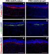
The images illustrate levels of profibrotic proteins in PRK-applied and AAV5-naked or AAV5-Smad7 treated eyes. Immunostaining for F-actin (A, B), α-SMA (C, D), and fibronectin (E, F), respectively. Scale Bar: 100 μm. (Reproduced from: Gupta S, Rodier JT, Sharma A, Giuliano EA, Sinha PR, Hesemann NP, Ghosh A, Mohan RR. (2017) Targeted AAV5-Smad7 gene therapy inhibits corneal scarring in vivo. PLoS One 2017;12:e0172928).
Fig. 3.
Fig. 3.
Dynamics of wound healing. A typical course of healing is presented for a c-met–transduced and vector-transduced cornea. rAV-vector–transduced diabetic cornea (top row) healed in 7 days, whereas rAV-met–transduced fellow cornea (bottom row) healed in 3 days. Pictures of live healing corneas are shown. Dashed line: shows the boundaries of the unhealed wound region. W, wound zone; E, migrating epithelium. (Reproduced from: Saghizadeh M, Kramerov AA, Yu FS, Castro MG, Ljubimov AV. Normalization of wound healing and diabetic markers in organ cultured human diabetic corneas by adenoviral delivery of c-Met gene. Invest Ophthalmol Vis Sci. 2010;51:1970–1980).
Fig. 4.
Fig. 4.
Normalizing effects of nanobioconjugates (NBCs) in diabetic limbal epithelial cell (LEC) cultures. A, By immunostaining, control LEC showed rare keratin (K)15-positive cells. After lead NBC treatment, the number of positive cells markedly increased and became similar to normal LEC. B, Lead NBC boosting c-Met expression by inhibiting miR-409 and blocking cathepsin F (CF) expression by the attached antisense oligonucleotides (AON) accelerated scratch wound healing in diabetic LEC (phase contrast). C, Lead NBC significantly reduced open scratch wound area compared to NBC with standard AON. The addition of a second NBC inhibiting MMP-10 (combo) further accelerated wound healing; however, the difference from lead NBC was not significant. The graph comprises data from four to five independent experiments using LECs from three independent cases. (Reproduced with permission from: Kramerov AA, Shah R, Ding H, et al. Novel nanopolymer RNA therapeutics normalize human diabetic corneal wound healing and epithelial stem cells. Nanomedicine. 2021;32:102332).
Fig. 5.
Fig. 5.
The appearance of the corneal graft 14 days after the operation. A, In group I (negative control; subconjunctival injection of saline after surgery), the graft showed oedema and new blood vessel growth into the centre of the graft. B, in the group II (IL-1ra plasmid gene injection into the corneal stroma before donor graft collection), the graft showed mild oedema, and fewer new blood vessels were observed than in controls. C-D, in the groups III (IL-1ra plasmid gene injection into the anterior chamber after graft-graft bed alignment suturing) and group IV (positive control; subconjunctival injection of IL-1ra protein after surgery), the graft was transparent, and no neovascularisation was found in the centre of the graft. (Reproduced from: Yuan J, Liu Y, Huang W, Zhou S, Ling S, Chen J. The experimental treatment of corneal graft rejection with the interleukin-1 receptor antagonist (IL-1ra) gene. PLoS One. 2013;8:e60714).
Fig. 6.
Fig. 6.
Effect of subconjunctival injection of rAAV-angiostatin on alkali burn-induced cornea neovascularization regression. One week before infliction of the alkali burn with 1 N NaOH in rats, the rats were assigned randomly either to a treatment group or control group. The treatment group received a subconjunctival injection of rAAV-angiostatin three weeks before the alkali burn, and the control group received blank rAAV viral vector treatment. Corneal NV was examined daily by a slit lamp for two weeks. Representative photographs of alkali burn-induced corneal NV at days 7 and 14are shown. A: Dense NV is growing toward the central cornea. Seven days after the alkali burn, there was severe corneal opacity and epithelial defects in the rAAV viral vector control eye. B: The rAAV-angiostatin injected eye seven days after the alkali burn is shown. Note the scarcity of corneal NV and the mild to moderate corneal opacity. C: The rAAV viral vector control eye at day 14 follow-up is shown. Corneal NV and pannus maturation were evident. D: The rAAV-angiostatin injected eye 14 days after the alkali burn is shown. Not only the corneal NV was absent, but also the central corneal opacity was mild. E: The corneal NV area (mean±SD, n=6) measured at seven days after exposure to the alkali burn is shown. The rAAV-angiostatin treated group demonstrated a significantly smaller NV area compared with the rAAV viral vector control group (the asterisk means that p<0.05). (Reproduced from: Cheng HC, Yeh SI, Tsao YP, Kuo PC. Subconjunctival injection of recombinant AAV-angiostatin ameliorates alkali burn-induced corneal angiogenesis. Mol Vis. 2007;13:2344–2352).
Fig. 7.
Fig. 7.
IOP of rabbits during a 28-d period treated with Ad-p27 vs. controls (PBS or mitomycin C, MMC) (n=6). Data are given as mean ± SD. bP<0.05, cP<0.01 vs control. (Reproduced with permission from: Yang JG, Sun NX, Cui LJ, Wang XH, Feng ZH. Adenovirus-mediated delivery of p27(KIP1) to prevent wound healing after experimental glaucoma filtration surgery. Acta Pharmacol Sin. 2009;30:413–423).

Similar articles

Cited by

References

    1. Anguela XM, High KA. Entering the modern era of gene therapy. Annu Rev Med 2019; 70: 273–88. 10.1146/annurev-med-012017-043332 - DOI - PubMed
    1. Wirth T, Parker N, Ylä-Herttuala S. History of gene therapy. Gene 2013; 525(2): 162–9. 10.1016/j.gene.2013.03.137 - DOI - PubMed
    1. Dunbar CE, High KA, Joung JK, Kohn DB, Ozawa K, Sadelain M. Gene therapy comes of age. Science 2018; 359(6372): eaan4672. 10.1126/science.aan4672 - DOI - PubMed
    1. High KA, Roncarolo MG. Gene Therapy. N Engl J Med 2019; 381(5): 455–64. 10.1056/NEJMra1706910 - DOI - PubMed
    1. Bennett J, Maguire AM. Gene therapy for ocular disease. Mol Ther 2000; 1(6): 501–5. 10.1006/mthe.2000.0080 - DOI - PubMed

Publication types