Case Reports
. 2021 Apr 26;21(1):215.
doi: 10.1186/s12893-021-01157-8.

Lumbar spinal stenosis combined with obesity-induced idiopathic spinal epidural lipomatosis treated with posterior lumbar fusion: case report

Affiliations
Case Reports

Lumbar spinal stenosis combined with obesity-induced idiopathic spinal epidural lipomatosis treated with posterior lumbar fusion: case report

Xiao Han et al. BMC Surg. .

Abstract

Background: Spinal epidural lipomatosis is a rare cause of lumbar spinal stenosis. While conservative therapy is applicable for most of cases, surgical intervention is necessary for severe ones. This is the first time we apply this modified technique to this disease.

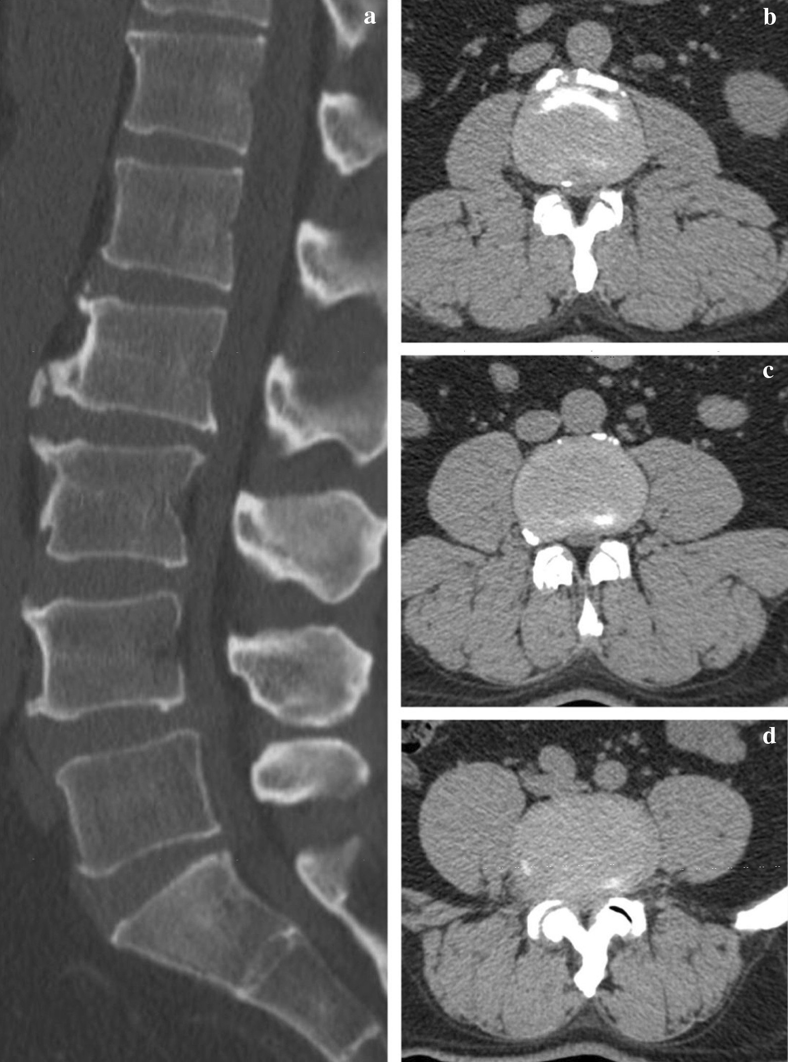
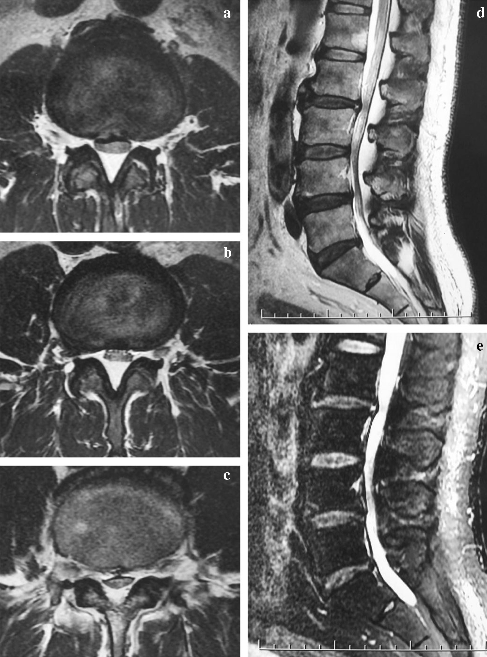
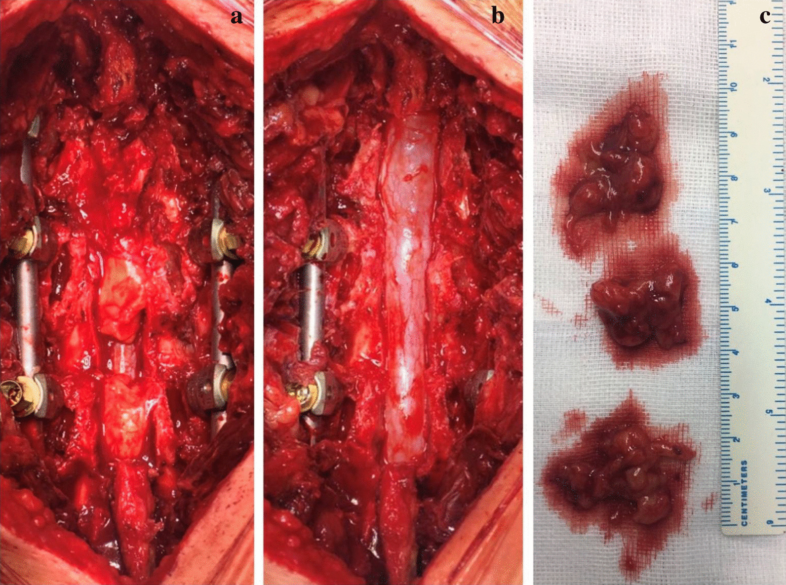
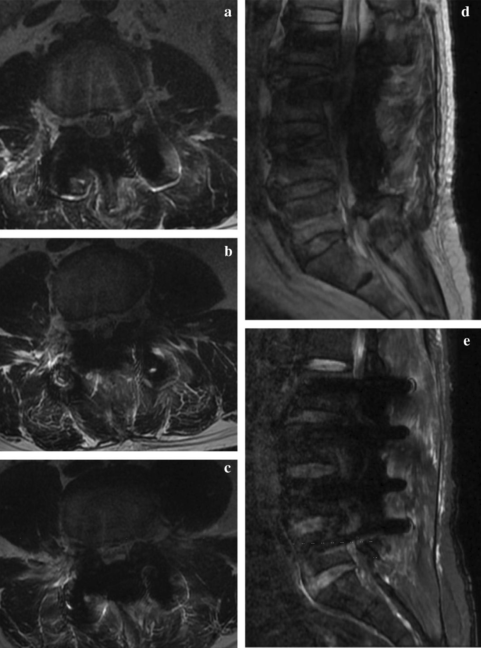
Case presentation: The case is a 53-year-old man. He is 175 cm tall and weighs 102 kg (body mass index 33.3 kg/cm2), presenting with low back pain and bilateral legs pain and numbness. Radiological examination showed severe lumbar spinal stenosis resulting from adipose hyperplasia, combined with hyperosteogeny and hypertrophy of ligaments, which are common etiological factors. Posterior decompression, internal fixation and a modified articular fusion technique was performed on this patient, and regular follow-up that up o 22 months showed outstanding clinical outcomes.

Conclusions: A suitable style of posterior lumbar fusion should be considered to especially severe case with lumbar spinal stenosis and idiopathic spinal epidural lipomatosis.

Keywords: Case report; Lumbar spinal stenosis; Obesity; Posterior lumbar fusion; Spinal epidural lipomatosis.


Conflict of interest statement

All authors declare that they have no competing interests.

Figures

Fig. 1
Fig. 1
Preoperative CT scan. Preoperative sagittal CT scan indicates intervertebral space narrow, vertebral body bone hyperplasia, “Y” shape homogenious hypodense shadow and oval compression of caudal spinal cord. Axial images shows stenosis frome L2 to L5 and mild hypertrophy of ligamentum flavum and facet joint. a Sagittal image. b Axial image of L2/3. c Axial image of L3/4. d Axial image of L4/5
Fig. 2
Fig. 2
Preoperative MRI. T2-weighted axial images show classic “Y-sign” thecal compression secondary to epidural lipomatosis from L2-L5. a Axial image of L2/3. b Axial image of L3/4. c Axial image of L4/5. d T2-weighted sagittal image shows epidural lipomatosis as short homogeneous signal, compression and ventral shift spinal cord. e Fat suppression image shows suppression of adipose tissue
Fig. 3
Fig. 3
Intraoperative image. a Intraoperative image shows accumulation of epidural adipose tissue. b Decompression range includes bony structure and adipose tissue. c Resected adipose tissue with normal appearance
Fig. 4
Fig. 4
Preoperative and immediate postoperative X-Ray. Immediate postoperative X-Ray image in comparison of the preoperative counterparts shows satisfactory location of graft. a Preoperative coronal image. b Preoperative sagittal image. c Postoperative coronal image. d Postoperative sagittal image
Fig. 5
Fig. 5
Immediate postoperative MRI. Immediate postoperative MRI image shows complete cancelation of extral adipose tissue and adequate decompression. a Axial image of L2/3. b Axial image of L3/4. c Axial image of L4/5. d T2-weighted sagittal image. e Fat suppression image
Fig. 6
Fig. 6
Follow-up X-Rays. Follow-up imaging examination shows consistent spinal stability and excellent internal fixation. a Coronal image of 3 months after surgery. b Sagittal image of 3 months after surgery. c Coronal image of 6 months after surgery. d Sagittal image of 6 months after surgery. e Coronal image of 22 months after surgery. f Sagittal image of 22 months after surgery
Fig. 7
Fig. 7
Postoperative CT scan and MRI. Postoperative CT scan shows satisfactory fusion rate of joint facet. MRI has not detected reoccurrence of spinal epidural lipomatosis. a Sagittal CT image of 6 months after surgery. b Sagittal CT image of 22 months after surgery. c Sagittal MRI image of 6 months after surgery. d Sagittal MRI image of 22 months after surgery

References

    1. Fassett DR, Schmidt MH. Spinal epidural lipomatosis: a review of its causes and recommendations for treatment. Neurosurg Focus. 2004;16(4):E11. - PubMed
    1. Fogel GR, Cunningham PY, 3rd, Esses SI. Spinal epidural lipomatosis: case reports, literature review and meta-analysis. Spine J. 2005;5(2):202–211. doi: 10.1016/j.spinee.2004.05.252. - DOI - PubMed
    1. Kotilainen E, Hohenthal U, Karhu J, Kotilainen P. Spinal epidural lipomatosis caused by corticosteroid treatment in ulcerative colitis. Eur J Intern Med. 2006;17(2):138–140. doi: 10.1016/j.ejim.2005.08.011. - DOI - PubMed
    1. Kim K, Mendelis J, Cho W. spinal epidural lipomatosis: a review of pathogenesis, characteristics, clinical presentation, and management. Glob Spine J. 2019;9(6):658–665. doi: 10.1177/2192568218793617. - DOI - PMC - PubMed
    1. Lee M, Lekias J, Gubbay SS, Hurst PE. Spinal cord compression by extradural fat after renal transplantation. Med J Aust. 1975;1(7):201–203. doi: 10.5694/j.1326-5377.1975.tb111328.x. - DOI - PubMed

Publication types