Review
. 2021 May;31(2):157-166.
doi: 10.1016/j.nic.2021.02.002.

Extracranial Vascular Disease: Carotid Stenosis and Plaque Imaging

Affiliations
Review

Extracranial Vascular Disease: Carotid Stenosis and Plaque Imaging

Hediyeh Baradaran et al. Neuroimaging Clin N Am. 2021 May.

Abstract

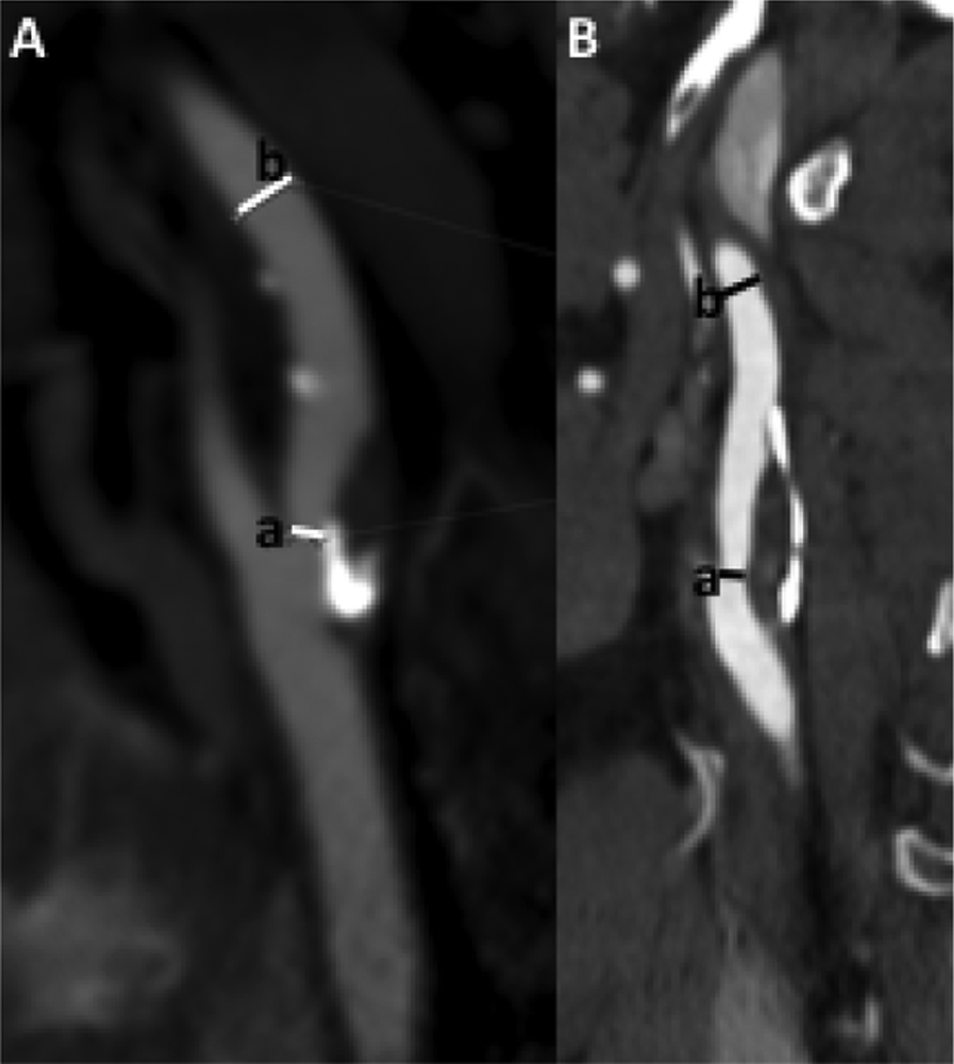
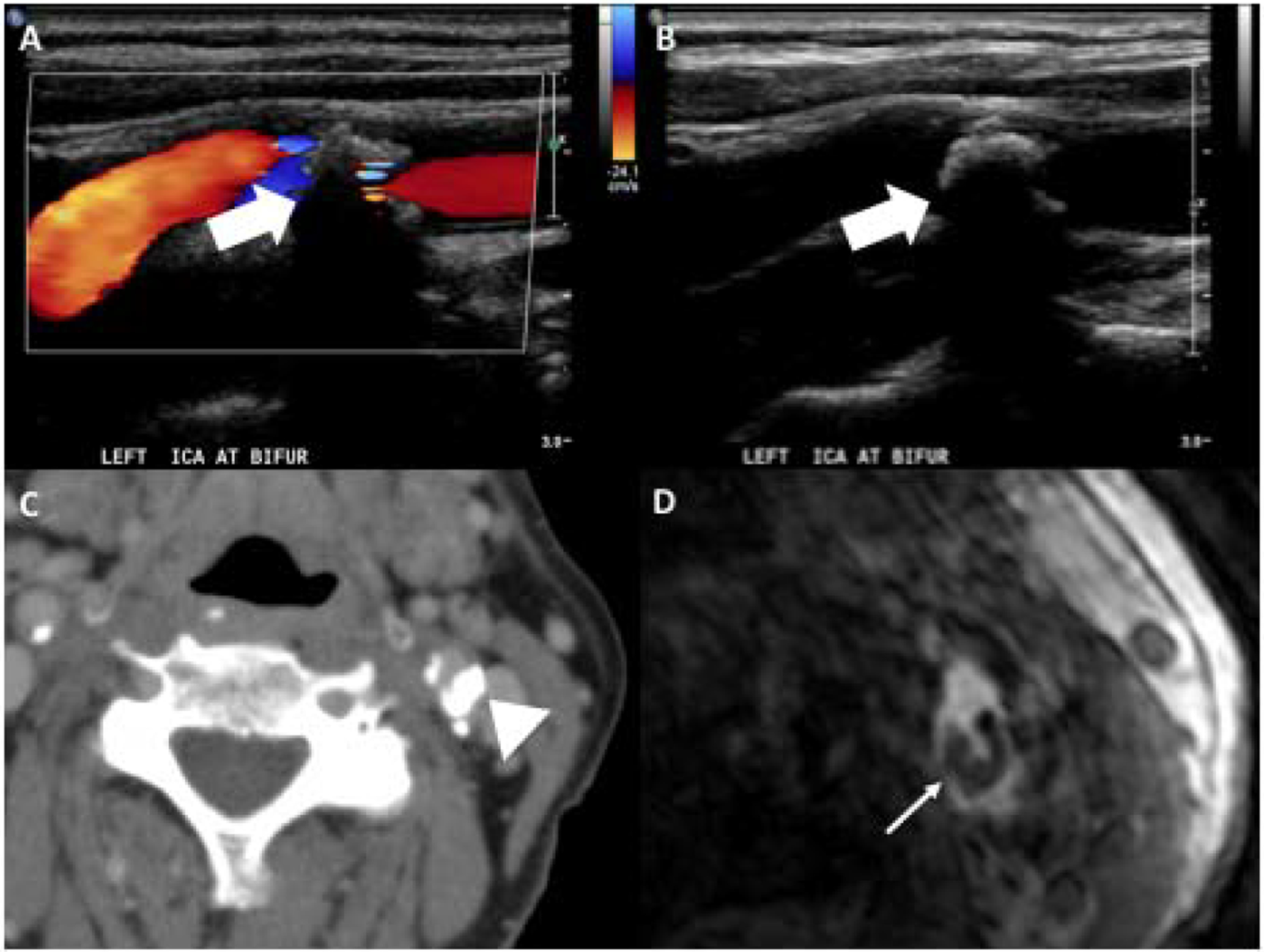
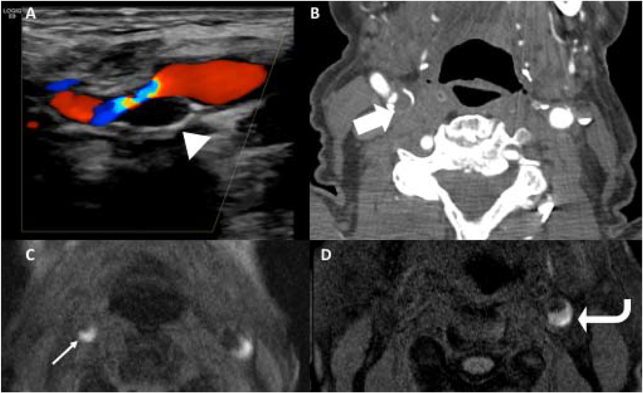
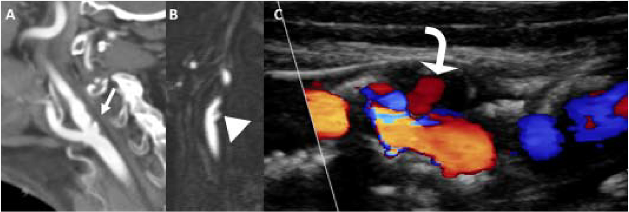
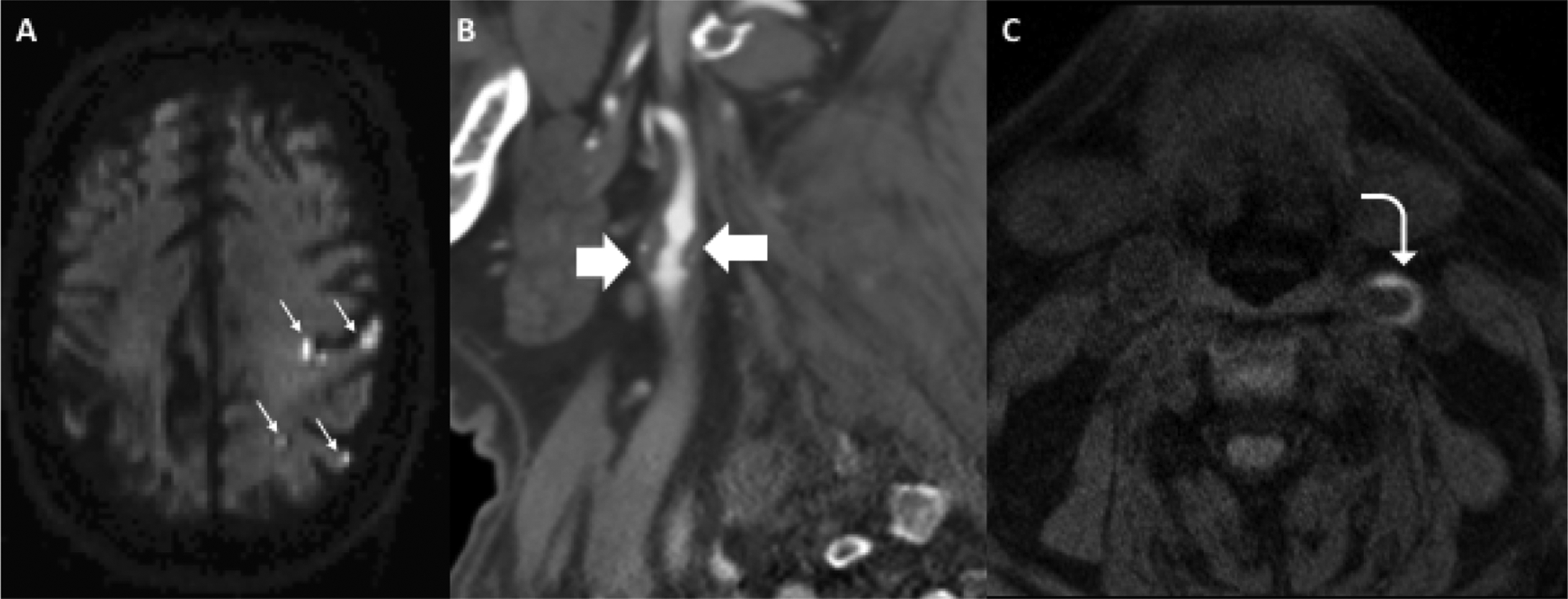
Carotid atherosclerosis is an important contributor to ischemic stroke. When imaging carotid atherosclerosis, it is essential to describe both the degree of luminal stenosis and specific plaque characteristics because both are risk factors for cerebrovascular ischemia. Carotid atherosclerosis can be accurately assessed using multiple imaging techniques, including ultrasonography, computed tomography angiography, and magnetic resonance angiography. By understanding the underlying histopathology, the specific plaque characteristics on each of these imaging modalities can be appreciated. This article briefly describes some of the most commonly encountered plaque features, including plaque calcification, intraplaque hemorrhage, lipid-rich necrotic core, and plaque ulceration.

Keywords: CT; Carotid atherosclerosis; Carotid plaque; Intraplaque hemorrhage; MR imaging.


Conflict of interest statement

Disclosure The authors have nothing to disclose relevant to the submitted work. Dr A. Gupta reports nonfinancial support from GE Healthcare and nonfinancial support from Siemens Medical Solutions USA, Inc, outside the submitted work.

Figures

Figure 1.
Figure 1.
Measuring stenosis by NASCET criteria involves first identifying and measuring the area of narrowest luminal diameter (a) and then measuring the luminal diameter in the normal more distal cervical ICA (b). These measurements should be made in plane with the vessel, accounting for vessel tortuosity. NASCET stenosis percentage equals (b-a)/b.
Figure 2.
Figure 2.
85-year-old female with large calcified plaque at the left carotid bifurcation as seen on US, CT, and MR. Doppler (A) and gray scale (B) US images demonstrate large echogenic plaque with posterior acoustic shadowing (white large arrows). The posterior acoustic shadowing limits appreciation of the size of the plaque. CT on the same patient (C) shows large calcified plaque in the posterior aspect of the left carotid bifurcation (arrowhead). Axial slice of 3D MR TOF (D) shows area of hypointensity in the posterior aspect of the left carotid bifurcation corresponding to the plaque calcification (narrow white arrow).
Figure 3.
Figure 3.
A-C show US, CTA, and axial 3D MPRAGE MR on the same patient. This 87-year-old female with acute right sided stroke (not pictured) had large echolucent plaque in the right carotid bifurcation on US (arrowhead), large soft/fibrofatty plaque on CTA (large block arrow), and crescentic T1 hyperintense plaque on 3D MPRAGE in the right (narrow white arrow) and left proximal internal carotid arteries (ICAs), consistent with intraplaque hemorrhage. D is another patient who presented with an acute left sided infarction with large crescentic T1 hyperintense plaque in the proximal left ICA (curved arrow), consistent with intraplaque hemorrhage. The signal is greater than 2x the intensity of adjacent sternocleidomastoid muscle.
Figure 4.
Figure 4.
Plaque ulceration. Sagittal reformations of CTA (A) and contrast-enhanced MRA (B) demonstrate large plaque ulcerations (white arrow and arrowhead) in the proximal ICAs in two different patients. Carotid US (C) demonstrating Doppler flow (curved arrow) within a large echolucent plaque in the proximal ICA, compatible with large plaque ulceration.
Figure 5.
Figure 5.
Case study of 71-year-old female presenting with acute right-sided weakness found to have multiple acute infarctions throughout the left cerebral hemisphere on axial DWI (A, small white arrows). Immediately after rapid brain MRI, she had a CTA head and neck which showed a large soft/fibrofatty plaque in the proximal left ICA (B, block arrows) with areas of ulceration resulting in less than 50% stenosis by NASCET criteria. On axial MPRAGE, she had areas of crescentic T1 hyperintensity (C, curved arrow) consistent with intraplaque hemorrhage.

References

    1. Sacco RL, Kasner SE, Broderick JP, et al. An Updated Definition of Stroke for the 21st Century. Stroke. 2013;44(7):2064. - PMC - PubMed
    1. Wityk R, Lehman D, Klag M, Coresh J, Ahn H, Litt B. Race and sex differences in the distribution of cerebral atherosclerosis. Stroke. 1996;27(11):1974–1980. - PubMed
    1. Rothwell P, Eliasziw M, Gutnikov S, et al. Analysis of pooled data from the randomised controlled trials of endarterectomy for symptomatic carotid stenosis. The Lancet. 2003;361(9352):107–116. - PubMed
    1. Group ECSTC. Randomised trial of endarterectomy for recently symptomatic carotid stenosis: final results of the MRC European Carotid Surgery Trial (ECST). The Lancet. 1998;351(9113):1379–1387. - PubMed
    1. Walker MD, Marler JR, Goldstein M, et al. Endarterectomy for asymptomatic carotid artery stenosis. Jama. 1995;273(18):1421–1428. - PubMed

MeSH terms