

Dual nature of human ACE2 glycosylation in binding to SARS-CoV-2 spike
- PMID: 33903171
- PMCID: PMC8126795
- DOI: 10.1073/pnas.2100425118
Dual nature of human ACE2 glycosylation in binding to SARS-CoV-2 spike
Abstract
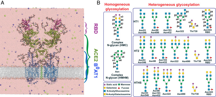
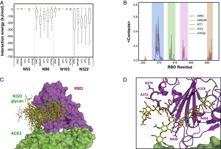
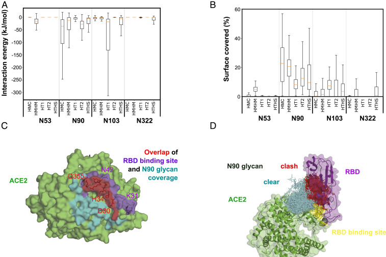
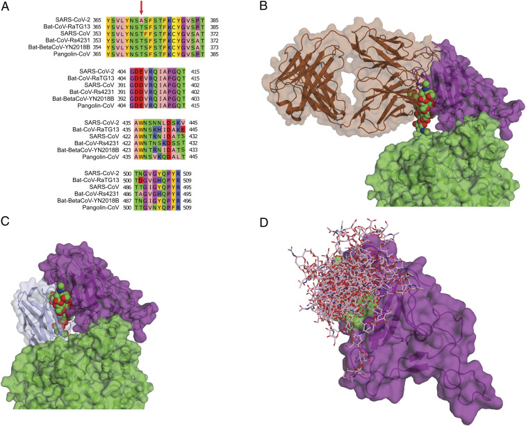
Binding of the spike protein of SARS-CoV-2 to the human angiotensin-converting enzyme 2 (ACE2) receptor triggers translocation of the virus into cells. Both the ACE2 receptor and the spike protein are heavily glycosylated, including at sites near their binding interface. We built fully glycosylated models of the ACE2 receptor bound to the receptor binding domain (RBD) of the SARS-CoV-2 spike protein. Using atomistic molecular dynamics (MD) simulations, we found that the glycosylation of the human ACE2 receptor contributes substantially to the binding of the virus. Interestingly, the glycans at two glycosylation sites, N90 and N322, have opposite effects on spike protein binding. The glycan at the N90 site partly covers the binding interface of the spike RBD. Therefore, this glycan can interfere with the binding of the spike protein and protect against docking of the virus to the cell. By contrast, the glycan at the N322 site interacts tightly with the RBD of the ACE2-bound spike protein and strengthens the complex. Remarkably, the N322 glycan binds to a conserved region of the spike protein identified previously as a cryptic epitope for a neutralizing antibody. By mapping the glycan binding sites, our MD simulations aid in the targeted development of neutralizing antibodies and SARS-CoV-2 fusion inhibitors.
Keywords: ACE2 receptor; SARS-CoV-2; glycosylation; molecular dynamics; virus-host interaction.
Copyright © 2021 the Author(s). Published by PNAS.
Conflict of interest statement
The authors declare no competing interest.
Figures
Similar articles
-
ACE2 glycans preferentially interact with SARS-CoV-2 over SARS-CoV.Chem Commun (Camb). 2021 Jun 15;57(48):5949-5952. doi: 10.1039/d1cc02305e. Chem Commun (Camb). 2021. PMID: 34019602
-
Inhibition of SARS-CoV-2 viral entry upon blocking N- and O-glycan elaboration.Elife. 2020 Oct 26;9:e61552. doi: 10.7554/eLife.61552. Elife. 2020. PMID: 33103998 Free PMC article.
-
S494 O-glycosylation site on the SARS-CoV-2 RBD affects the virus affinity to ACE2 and its infectivity; a molecular dynamics study.Sci Rep. 2021 Jul 26;11(1):15162. doi: 10.1038/s41598-021-94602-w. Sci Rep. 2021. PMID: 34312429 Free PMC article.
-
Interactions of angiotensin-converting enzyme-2 (ACE2) and SARS-CoV-2 spike receptor-binding domain (RBD): a structural perspective.Mol Biol Rep. 2023 Mar;50(3):2713-2721. doi: 10.1007/s11033-022-08193-4. Epub 2022 Dec 23. Mol Biol Rep. 2023. PMID: 36562937 Free PMC article. Review.
-
Structural basis of severe acute respiratory syndrome coronavirus 2 infection.Curr Opin HIV AIDS. 2021 Jan;16(1):74-81. doi: 10.1097/COH.0000000000000658. Curr Opin HIV AIDS. 2021. PMID: 33186231 Review.
Cited by
-
Structural remodeling of SARS-CoV-2 spike protein glycans reveals the regulatory roles in receptor-binding affinity.Glycobiology. 2023 Mar 6;33(2):126-137. doi: 10.1093/glycob/cwac077. Glycobiology. 2023. PMID: 36370046 Free PMC article.
-
Structural and Functional Characterization of SARS-CoV-2 RBD Domains Produced in Mammalian Cells.Anal Chem. 2021 May 4;93(17):6839-6847. doi: 10.1021/acs.analchem.1c00893. Epub 2021 Apr 19. Anal Chem. 2021. PMID: 33871970 Free PMC article.
-
BPP: a platform for automatic biochemical pathway prediction.Brief Bioinform. 2024 Jul 25;25(5):bbae355. doi: 10.1093/bib/bbae355. Brief Bioinform. 2024. PMID: 39082653 Free PMC article.
-
COVID-19 and the eye: alternative facts The 2022 Bowman Club, David L. Easty lecture.BMJ Open Ophthalmol. 2022 May;7(1):e001042. doi: 10.1136/bmjophth-2022-001042. BMJ Open Ophthalmol. 2022. PMID: 35675203 Free PMC article. Review.
-
Expression pattern and function of SARS-CoV-2 receptor ACE2.Biosaf Health. 2021 Dec;3(6):312-318. doi: 10.1016/j.bsheal.2021.08.003. Epub 2021 Aug 27. Biosaf Health. 2021. PMID: 34466800 Free PMC article. Review.
References
-
- Donoghue M., et al. ., A novel angiotensin-converting enzyme-related carboxypeptidase (ACE2) converts angiotensin I to angiotensin 1-9. Circ. Res. 87, E1–E9 (2000). - PubMed
Publication types
MeSH terms
Substances
LinkOut - more resources
Full Text Sources
Other Literature Sources
Molecular Biology Databases
Miscellaneous
