. 2021 Jul;45(7):1565-1575.
doi: 10.1038/s41366-021-00820-7. Epub 2021 Apr 26.

GHS-R suppression in adipose tissues protects against obesity and insulin resistance by regulating adipose angiogenesis and fibrosis

Affiliations

GHS-R suppression in adipose tissues protects against obesity and insulin resistance by regulating adipose angiogenesis and fibrosis

Jong Han Lee et al. Int J Obes (Lond). 2021 Jul.

Abstract

Background/objectives: Ghrelin is an orexigenic hormone that increases food intake, adiposity, and insulin resistance through its receptor Growth Hormone Secretagogue Receptor (GHS-R). We previously showed that ghrelin/GHS-R signaling has important roles in regulation of energy homeostasis, and global deletion of GHS-R reduces obesity and improves insulin sensitivity by increasing thermogenesis. However, it is unknown whether GHS-R regulates thermogenic activation in adipose tissues directly.

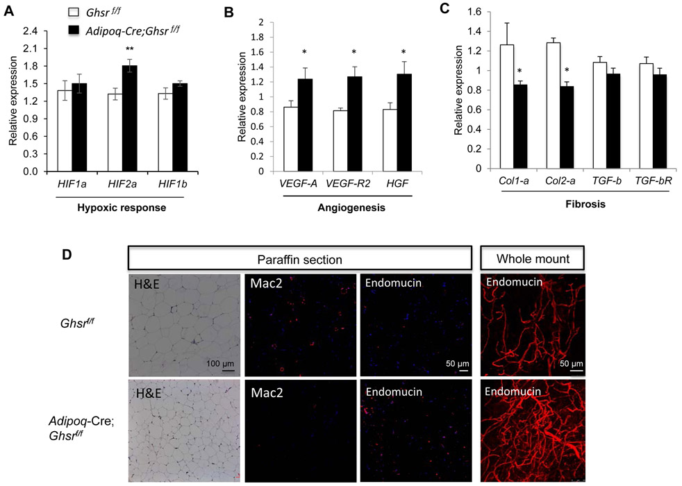
Methods: We generated a novel adipose tissue-specific GHS-R deletion mouse model and characterized the mice under regular diet (RD) and high-fat diet (HFD) feeding. Body composition was measured by Echo MRI. Metabolic profiling was determined by indirect calorimetry. Response to environmental stress was assessed using a TH-8 temperature monitoring system. Insulin sensitivity was evaluated by glucose and insulin tolerance tests. Tissue histology was analyzed by hematoxylin/eosin and immunofluorescent staining. Expression of genes involved in thermogenesis, angiogenesis and fibrosis in adipose tissues were analyzed by real-time PCR.

Results: Under RD feeding, adipose tissue-specific GHS-R deletion had little or no impact on metabolic parameters. However, under HFD feeding, adipose tissue-specific GHS-R deletion attenuated diet-induced obesity and insulin resistance, showing elevated physical activity and heat production. In addition, adipose tissue-specific GHS-R deletion increased expression of master adipose transcription regulator of peroxisome proliferator-activated receptor (PPAR) γ1 and adipokines of adiponectin and fibroblast growth factor (FGF) 21; and differentially modulated angiogenesis and fibrosis evident in both gene expression and histological analysis.

Conclusions: These results show that GHS-R has cell-autonomous effects in adipocytes, and suppression of GHS-R in adipose tissues protects against diet-induced obesity and insulin resistance by modulating adipose angiogenesis and fibrosis. These findings suggest adipose GHS-R may constitute a novel therapeutic target for treatment of obesity and metabolic syndrome.


Conflict of interest statement

Conflict of interest statement: The authors declare no conflict of interests.

Figures

Figure. 1.
Figure. 1.. Validation of Adipoq-Cre;Ghsrf/f mouse model and adipose tissue-specific GHS-R deletion reduced body weight and fat mass under HFD-feeding.
(A) Relative GHS-R gene expression in hypothalamus (Hypo), epididymal white adipose tissue (Epi), brown adipose tissue (BAT), inguinal white adipose tissue (Ingu), and muscle. n=5-8. *, p<0.05, Ghsrf/f vs. Adipoq-Cre;Ghsrf/f. (B) Body weight and (C) Fat percentage at 6-32 weeks of age. (D) The representative images of mouse, liver, epiWAT, and BAT. n=6-8. *, p<0.05, Ghsrf/f vs. Adipoq-Cre;Ghsrf/f.
Figure. 2.
Figure. 2.. HFD-fed adipose tissue-specific GHS-R deletion improved insulin sensitivity.
Metabolic profiles were performed using indirect calorimetry at 15-16 weeks of age; data under fed conditions present an average of last 3 days, and mice were fasted for 24 h before termination. (A) Food intake, (B) Physical activity, (C) Energy Expenditure (Heat production), (D) Resting metabolic rate (RMR), and (E) Respiratory exchange ratio (RER). n=5. *, p<0.05, Ghsrf/f vs. Adipoq-Cre;Ghsrf/f. (F) Blood glucose levels during GTT at 17 weeks of age. (G) Blood glucose levels during ITT at 22 weeks of age. n=6-8. *, p<0.05, Ghsrf/f vs. Adipoq-Cre;Ghsrf/f.
Figure. 3.
Figure. 3.. Adipose tissue-specific GHS-R deletion had no effect on thermogenesis.
HFD-fed mice at 32 weeks of age were individually caged at 4°C and provided with free access to food and water. (A) Rectal temperature was recorded hourly for 4 h. (B) Expression of thermogenic genes in BAT. (C) Expression of thermogenic genes in iWAT. n=5-6. *, p<0.05, Ghsrf/f vs. Adipoq-Cre;Ghsrf/f.
Figure. 4.
Figure. 4.. Adipose tissue-specific GHS-R deletion did not affect lipogenesis but increased transcription factor PPARγ1, and adipokines of FGF21 and adiponectin in epiWAT.
(A) Expression of lipogenic genes. (B) Expression of master adipose transcription factors PPARγ1 and PPARγ2. (C) Expression of adipokines of FGF21, adiponectin and leptin. n=5. *, p<0.05, Ghsrf/f vs. Adipoq-Cre;Ghsrf/f.
Figure. 5.
Figure. 5.. Adipose tissue-specific GHS-R deletion modulated angiogenesis and fibrosis in epiWAT under HFD feeding.
(A) Expression of hypoxia-inducible transcription factors. (B) Expression of angiogenic genes. (C) Expression of fibrotic genes. (D) H&E staining, macrophages marker Mac2 (red), and vasculature marker endomucin (red) staining in paraffin sections; endomucin staining in tissue whole mount, showing decreased macrophage infiltration and increased microvasculature in epiWAT of HFD-fed Adipoq-Cre;Ghsrf/f mice. n=5. *, p<0.05, Ghsrf/f vs. Adipoq-Cre;Ghsrf/f.
Figure. 6.
Figure. 6.. Schematic diagram of the proposed actions of GHS-R in adipose tissue under obesity.
Under obesity conditions, GHS-R inhibition in adipocytes mitigates adiposity and improves insulin sensitivity by the following potential mechanisms: 1) Activating transcriptional regulator PPARγ1 to enhance healthy adipokines of FGF21 and adiponectin, which decreases macrophage infiltration to reduce inflammation and Col1α/Col2α expression to suppress fibrosis; 2) Stimulating hypoxia transcription regulator HIF2α to increase the expression of VEGF, VEGF-R2, and HGF to promote angiogenesis. Together, these functional signaling cascades lead to reduced adiposity and improved insulin sensitivity.

Similar articles

Cited by

References

    1. Kursawe R, Caprio S, Giannini C, Narayan D, Lin A, D'Adamo E, et al.: Decreased transcription of ChREBP-alpha/beta isoforms in abdominal subcutaneous adipose tissue of obese adolescents with prediabetes or early type 2 diabetes: associations with insulin resistance and hyperglycemia. Diabetes 2013, 62:837–844. - PMC - PubMed
    1. Lee YH, Mottillo EP, Granneman JG: Adipose tissue plasticity from WAT to BAT and in between. Biochim Biophys Acta 2014, 1842:358–369. - PMC - PubMed
    1. Kozak LP, Koza RA, Anunciado-Koza R: Brown fat thermogenesis and body weight regulation in mice: relevance to humans. Int J Obes (Lond) 2010, 34 Suppl 1:S23–27. - PubMed
    1. Lin L, Lee JH, Bongmba OY, Ma X, Sheikh-Hamad D, Sun Y: The suppression of ghrelin signaling mitigates age-associated thermogenic impairment. Aging (Albany NY) 2014, 6:1019–1032. - PMC - PubMed
    1. Sacks H, Symonds ME: Anatomical locations of human brown adipose tissue: functional relevance and implications in obesity and type 2 diabetes. Diabetes 2013, 62:1783–1790. - PMC - PubMed

Publication types