

Faster Lumbar Spine Bone Loss in Midlife Predicts Subsequent Fracture Independent of Starting Bone Mineral Density
- PMID: 33903908
- PMCID: PMC8208668
- DOI: 10.1210/clinem/dgab279
Faster Lumbar Spine Bone Loss in Midlife Predicts Subsequent Fracture Independent of Starting Bone Mineral Density
Abstract
Context: Bone mineral density (BMD) decreases rapidly during menopause transition (MT), and continues to decline in postmenopause.
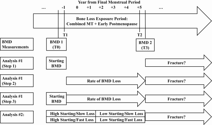
Objective: This work aims to examine whether faster BMD loss during the combined MT and early postmenopause is associated with incident fracture, independent of starting BMD, before the MT.
Methods: The Study of Women's Health Across the Nation, a longitudinal cohort study, included 451 women, initially premenopausal or early perimenopausal, and those transitioned to postmenopause. Main outcome measures included time to first fracture after early postmenopause.
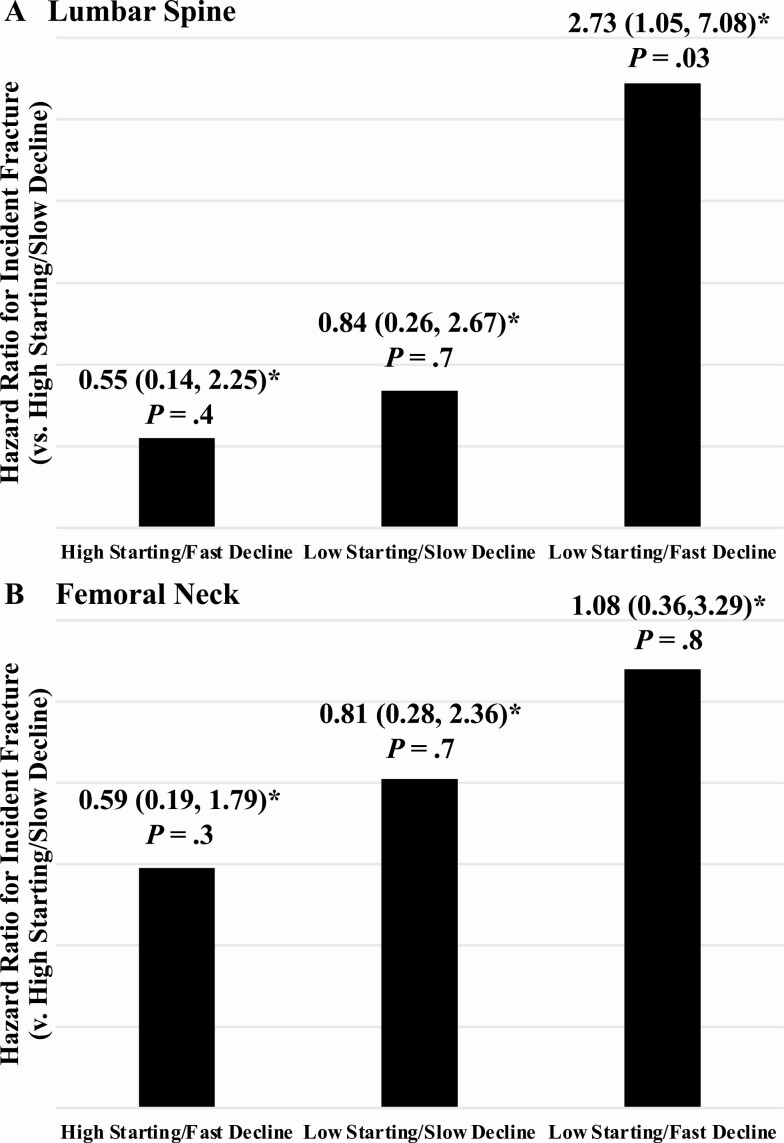
Results: In Cox proportional hazards regression, adjusted for age, body mass index, race/ethnicity, study site, use of vitamin D and calcium supplements, and use of bone-detrimental or -beneficial medications, each SD decrement in lumbar spine (LS) BMD before MT was associated with a 78% increment in fracture hazard (P = .007). Each 1% per year faster decline in LS BMD was related to a 56% greater fracture hazard (P = .04). Rate of LS BMD decline predicted future fracture, independent of starting BMD. Women with a starting LS BMD below the sample median, and an LS BMD decline rate faster than the sample median had a 2.7-fold greater fracture hazard (P = .03). At the femoral neck, neither starting BMD nor rate of BMD decline was associated with fracture.
Conclusion: At the LS, starting BMD before the MT and rate of decline during the combined MT and early postmenopause are independent risk factors for fracture. Women with a below-median starting LS BMD and a faster-than-median LS BMD decline have the greatest fracture risk.
Keywords: bone mineral density; fracture; general population studies; menopause.
© The Author(s) 2021. Published by Oxford University Press on behalf of the Endocrine Society. All rights reserved. For permissions, please e-mail: journals.permissions@oup.com.
Figures
References
-
- Burger H, van Daele PL, Algra D, et al. . The association between age and bone mineral density in men and women aged 55 years and over: the Rotterdam Study. Bone Miner. 1994;25(1):1-13. - PubMed
-
- Ravn P, Hetland ML, Overgaard K, Christiansen C. Premenopausal and postmenopausal changes in bone mineral density of the proximal femur measured by dual-energy X-ray absorptiometry. J Bone Miner Res. 1994;9(12):1975-1980. - PubMed
-
- Warming L, Hassager C, Christiansen C. Changes in bone mineral density with age in men and women: a longitudinal study. Osteoporos Int. 2002;13(2):105-112. - PubMed
Publication types
MeSH terms
Grants and funding
LinkOut - more resources
Full Text Sources
Other Literature Sources
Medical
