. 2021 Jul;28(4):e12691.
doi: 10.1111/xen.12691. Epub 2021 Apr 26.

Mixed xenogeneic porcine chimerism tolerizes human anti-pig natural antibody-producing cells in a humanized mouse model

Affiliations

Mixed xenogeneic porcine chimerism tolerizes human anti-pig natural antibody-producing cells in a humanized mouse model

Elizabeth E Waffarn et al. Xenotransplantation. 2021 Jul.

Abstract

Background: A major obstacle to the success of organ transplantation from pigs to humans, necessitated by the shortage of human organs, is robust humoral immune rejection by pig-reactive human antibodies. Mixed xenogeneic hematopoietic chimerism induces xenoreactive B cell tolerance in rodents, but whether mixed pig/human chimerism could induce tolerance of human B cells to pig xenoantigens is unknown.

Methods: We investigated this question using a humanized mouse model in which durable mixed (pig-human) xenogeneic chimerism can be established.

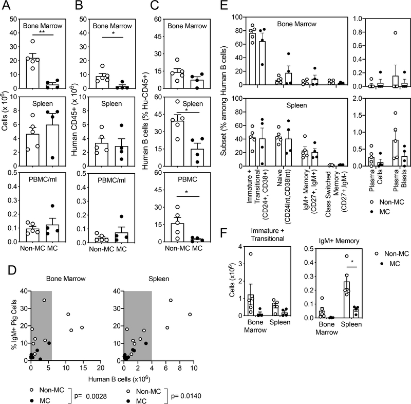
Results: Human natural anti-pig cytotoxic antibodies, predominantly IgM, are detectable in non-chimeric humanized mouse serum, and pig-reactive antibodies were reduced in mixed chimeric versus non-chimeric humanized mice. This difference required persistent mixed chimerism and was not due to the adsorption of antibodies on pig cells in vivo. Furthermore, human B cells from spleens of mixed chimeric mice produced lower levels of anti-pig antibodies when stimulated in vitro compared with those from non-chimeric mice.

Conclusions: Our findings demonstrate that mixed chimerism reduces human natural antibodies to pig xenoantigens, providing the first in vivo evidence of human B cell tolerance induction by mixed xenogeneic chimerism and supporting further evaluation of this approach for inducing human B cell tolerance to xenografts.

Keywords: B cells; Mixed Chimerism; humanized mice; xenoantibody; xenotolerance.


Conflict of interest statement

Disclosure: The authors declare no competing financial interests.

Figures

Figure 1.
Figure 1.. Natural cytotoxic anti-pig antibodies are detected in non-mixed chimeric humanized mice.
(A) Generation of humanized mice by injection of human fetal liver-derived CD34+ cells to sublethally irradiated adult NSG mice. (B) Levels of human IgM and IgG in humanized mouse serum, monitored by ELISA at 4-week intervals, n=4. (C-D) Presence of anti-pig antibodies determined by complement-dependent cytotoxicity assay. (C) Representative FCM plots showing cytotoxicity against pig PBMCs incubated with (from left) absence of serum or complement, complement but not serum, serum from a NHP sensitized against a pig skin graft (n=1) plus complement (as a positive control), commercial pooled human serum (n=1) plus complement, and humanized mouse serum plus complement. (D) Cytotoxicity against pig PBMCs mediated by sensitized NHP serum (n=1), commercial pooled human serum (n=1), or serum from humanized mice 15–20 weeks post-human CD34+ cell transplantation (n=22). Data represent at least two consistent (C, D) experiments.
Figure 2.
Figure 2.. Humanized mouse serum anti-pig responses are mediated by human IgM.
(A) To determine the isotypes of human anti-pig antibodies present in humanized mouse serum, target pig PBMCs were incubated with humanized mouse serum, followed by incubation with anti-human IgM-FITC and IgG-PE secondary antibodies. Representative FCM plots of isotypes of human antibodies binding the surface of target pig cells incubated with (from left) absence of serum or secondary antibodies, secondary antibodies only, positive control serum from a sensitized NHP (as in Figure 1) plus secondary antibodies, commercial pooled human serum (n=1) plus secondary antibodies, and humanized mouse serum plus secondary antibodies (right panel). (B) Percentages of pig PBMCs bound by IgM and IgG present in positive serum from a sensitized NHP (n=1) and commercial pooled human serum (n=1). Values shown were adjusted by subtracting the appropriate Ig binding frequency observed in pig PBMCs incubated with secondary antibodies alone from those of pig PBMCs incubated with both serum and secondary antibodies. (C) Percentages of pig PBMCs bound by human IgM or IgG following incubation with serum from humanized mice (n=7) demonstrating high anti-pig cytotoxicity 15–20 weeks post-human CD34+ cell transplantation, as described in Figure 1C-D. **p<0.001, Student’s paired t-test. Data from one of three experiments with similar results are shown.
Figure 3.
Figure 3.. Mixed xenogeneic chimerism results in decreased anti-pig antibodies in serum.
(A) Mixed chimeric (MC) mice were generated by injection of Gal+ pig BMCs and human fetal liver CD34+ cells to sublethally irradiated adult PCT-NSG mice. 15–20 weeks post-cell transfer, serum anti-pig responses from MC mice were compared with Non-MC humanized mice, using target pig PBMCs obtained from the same pig whose BMCs were used to generate the MC mice in each experiment. Mice from both groups were not thymectomized. (B) Cytotoxicity against pig cells mediated by serum of Non-MC and MC mice as determined by complement-dependent cytotoxicity assay. *** p<0.001 Mann-Whitney unpaired t test. Upper statistics; numbers and percentages of mice in each group in which anti-pig antibodies were detected. **p<0.01, Chi-square test, n=19–25. Data shown are pooled from 4 experiments. (C) Percentages of pig PBMCs bound by human IgM or IgG by FCM after incubation with serum from Non-MC and MC mice. ****p<0.01, two-way ANOVA with Tukey’s multiple comparison’s test. Data shown are from two pooled experiments. (D) Distribution of serum cytotoxicity against pig cells and concentration of total human IgM in serum from Non-MC and MC mice shown in (B). (E) Distribution of levels of anti-pig human IgM and concentration of total human IgM in serum for Non-MC and MC mice shown in (C). Mouse numbers are indicated on each figure.
Figure 4.
Figure 4.. Pig cells in MC mice are not bound by anti-pig natural antibodies in vivo.
To determine whether anti-pig natural antibodies are adsorbed onto pig cells in vivo, cells from BM, spleen, liver, peritoneal cavity lavage and PBMCs of MC mice generated from Gal+ pig BMC donors were stained with anti-human IgM. (A) Representative FCM plots from a MC mouse showing staining for surface human IgM among pig cells (top panels) and human B cells (bottom panels) in comparison to fluorescence minus one (FMO) controls lacking only human IgM staining (right panels). (B) Frequency of IgM+ pig cells compared to FMO controls among tissues from MC mice, n=5. Student’s t-test. Data represent four experiments with consistent results.
Figure 5.
Figure 5.. Reduced B cell reconstitution in MC mice.
Human cell reconstitution of Non-MC and MC mice generated from Gal+ pig BMC donors at 16–20 weeks post transplantation. Tissue cell number (A), Human CD45+ cell numbers (B), and Human CD19+ cell frequency among human CD45+ cells (C). (D) Distribution of human B cell reconstitution (by number of CD19+ cells) with serum levels of human pig-reactive IgM antibodies assayed by pig cell binding in Non-MC and MC mice. P value represents Mann-Whitney test of serum anti-pig IgM binding among MC and Non-MC mice with similar B cell levels (within shaded range). (E) Phenotype of human B cells in the bone marrow and spleen by subset frequency (E) or number (F), as gated in Supplemental Figure 1. *p<0.05, Student’s t-test. Data shown represent one of two experiments with consistent results (A-C, E-F) (n=4–5 mice per group) or pooled results from both experiments (D).
Figure 6.
Figure 6.. Reduced anti-pig antibody production in vitro by splenic human B cells from MC mice.
Human CD45+ cells were isolated from the spleen 16 weeks after transplantation, plated at equal concentrations with regard to CD19+ human B cells, and cultured in the presence of anti-CD40 antibody, CpG-DNA, IL-10, and IL-2 in the absence of human serum. After 7 days, concentrated supernatants were assayed for reactivity against either Gal+ or Gal- target pig BMCs from the same source pig as used to construct MC mice. (A) Concentrations of human IgM or IgG secreted into human cell culture supernatants from Non-MC (n=14) and MC (n=9) mice are shown. (B) Percent binding of target pig BMCs by human IgM from supernatants of Non-MC (n=6) compared to MC mice (n=5). *p<0.05, Mann Whitney test, n=5–6. (C) Cytotoxicity mediated against pig BMCs by supernatants from Non-MC mice (n=10) and MC mice (n=7 Gal-, n=2 Gal+). *p<0.05, Mann Whitney test. Data shown in (A) and (C) were pooled from three experiments. Non-MC: Non-mixed chimeric mice; MC: mixed chimeric mice generated with Gal- or Gal+ pig BMCs as noted.
Figure 7.
Figure 7.. Antigen persistence is required to maintain reduced anti-pig B cell responses in MC mice.
(A) To evaluate whether durable pig chimerism was required for the reduction in anti-pig natural antibody responses in MC mice, MC mice generated from Gal+ pig BMC donors were treated weekly for 4 weeks prior to sacrifice with 74.11.10, a mouse monoclonal anti-pig class I MHC antibody to deplete pig cells (depleted MC, triangles). Depleted MC mice were an additional group included in the same experiments presented in Figure 3–5, but data shown here focus on the comparison between pig cell-depleted MC or un-depleted MC mice. Un-depleted MC mice received no injection. (B) Pig cell chimerism depicted representative FCM plots showing pig chimerism in depleted compared to undepleted MC mice and as pig cell frequencies among live cells in tissues at the time of euthanasia, n=4–5 mice, one of two representative experiments. (C) Levels of serum anti-pig antibodies evaluated by percentages of human IgM+ or IgG+ target pig PBMCs after incubation with serum from MC and MC-depleted mice. **p<0.01, Student t-test, n=9–12 mice pooled from 2 experiments. (D) Levels of total serum human IgM, n=9–12 mice pooled from 2 experiments. (E) Lack of correlation of serum IgM concentration and levels of anti-pig human IgM in serum, n=9–10 mice pooled from 2 experiments.

References

    1. Yamada K, Sykes M, Sachs DH. Tolerance in xenotransplantation. Curr Opin Organ Transplant. 2017;22(6):522–528. - PMC - PubMed
    1. Griesemer A, Yamada K, Sykes M. Xenotransplantation: immunological hurdles and progress toward tolerance. Immunol Rev. 2014;258(1):241–258. - PMC - PubMed
    1. Galili U Anti-Gal in Humans and Its Antigen the α-Gal Epitope. In: The Natural Anti-Gal Antibody As Foe Turned Friend In Medicine.2018:3–22.
    1. Lai L, Kolber-Simonds D, Park KW, et al.Production of alpha-1,3-galactosyltransferase knockout pigs by nuclear transfer cloning. Science. 2002;295(5557):1089–1092. - PubMed
    1. Phelps CJ, Koike C, Vaught TD, et al.Production of alpha 1,3-galactosyltransferase-deficient pigs. Science. 2003;299(5605):411–414. - PMC - PubMed

Publication types

Substances