

Cryofibrinogen-associated glomerulonephritis accompanied by advanced gastric cancer
- PMID: 33905105
- PMCID: PMC8494845
- DOI: 10.1007/s13730-021-00602-0
Cryofibrinogen-associated glomerulonephritis accompanied by advanced gastric cancer
Abstract
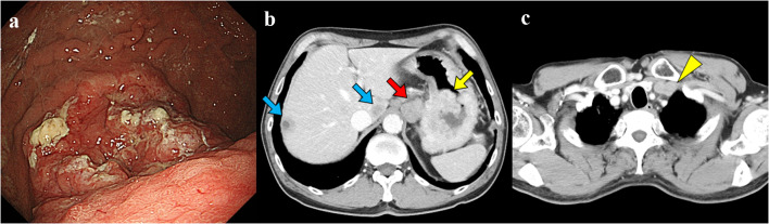
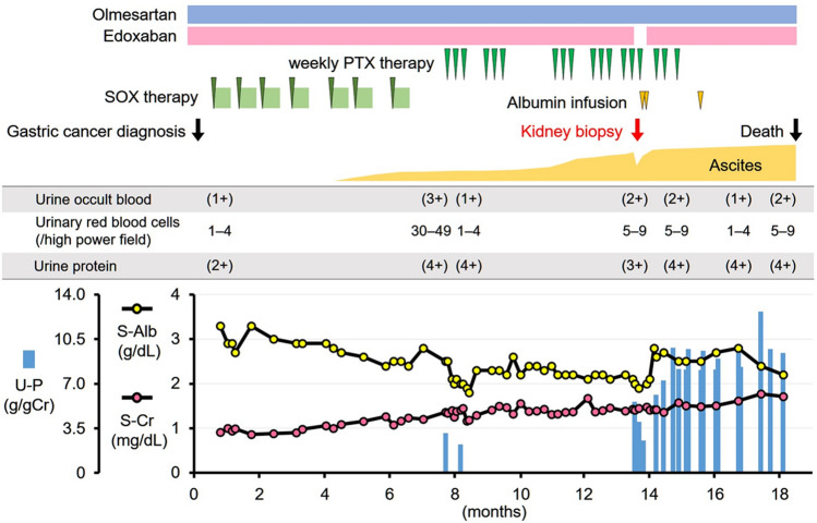
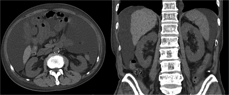
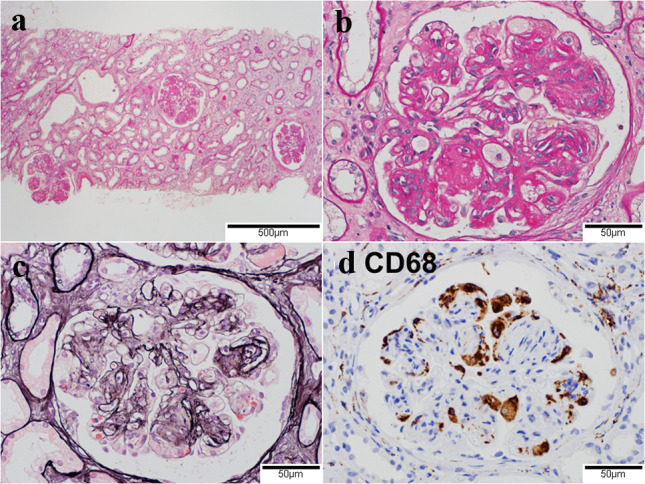
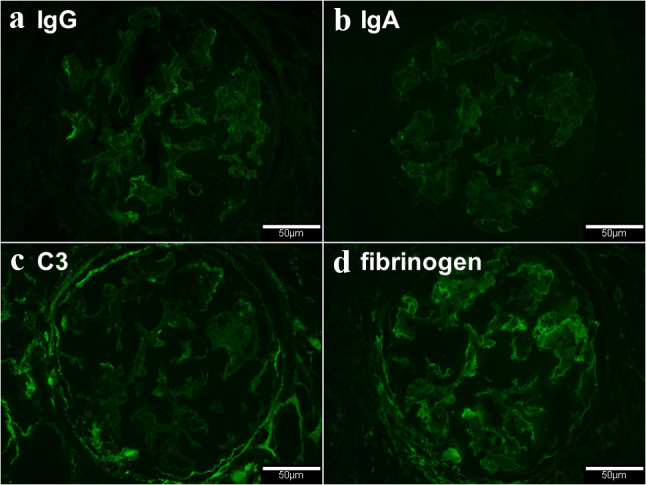
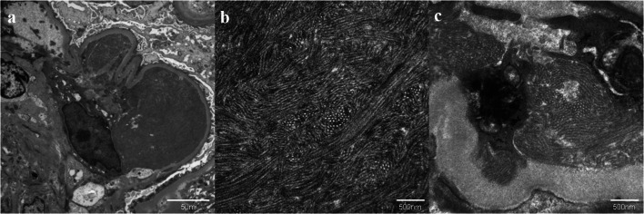
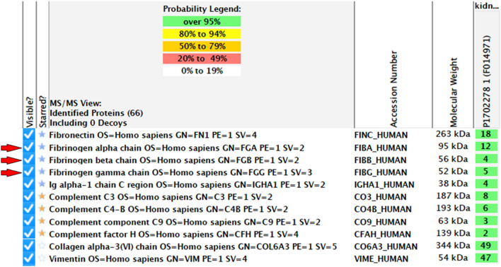
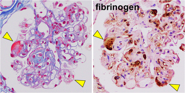
We had a 72-year-old man with advanced gastric cancer, poorly differentiated adenocarcinoma, receiving chemotherapy with S-1 (tegafur, gimeracil, and oteracil potassium) plus oxaliplatin. Ascites developed despite remission of gastric cancer and metastasis. Given no malignant cells in ascites, leg edema, renal impairment, hypoalbuminemia, and massive proteinuria, we diagnosed as nephrotic syndrome with microscopic hematuria. Renal biopsy showed membranoproliferative glomerulonephritis with no deposition of immunoglobulins and complements. Of note, electronic microscopy found organized deposits with microtubular structures in the glomerular capillary lumens and subendothelial spaces. The liquid chromatography-tandem mass spectrometry method detected fibrinogen alpha chain, beta chain, gamma chain, and fibronectin, and we eventually diagnosed cryofibrinogen-associated glomerulonephritis. Cryofibrinogen was not detected in plasma. He was expired at 5 months following renal biopsy due to the progression of refractory nephrotic syndrome. In addition to the detailed assessment of specifically organized deposits, the analysis using liquid chromatography-tandem mass spectrometry method is useful to diagnose cryofibrinogen-associated glomerulonephritis. We should consider cryofibrinogen-associated glomerulonephritis as a differential diagnosis when the patients with malignancy showed abnormal urinalysis and renal impairment, though it is a rare disease.
Keywords: LC–MS/MS; Mass spectrometry; Membranoproliferative glomerulonephritis; Nephrotic syndrome; Organized deposit.
© 2021. Japanese Society of Nephrology.
Conflict of interest statement
The authors have declared that no conflict of interest exists.
Figures
Similar articles
-
Discontinuing Hemodialysis through Corticosteroid Treatment in a Patient with Cryofibrinogen-associated Glomerulonephritis.Intern Med. 2024 Jul 1;63(13):1899-1905. doi: 10.2169/internalmedicine.2897-23. Epub 2023 Nov 6. Intern Med. 2024. PMID: 37926528 Free PMC article.
-
Characteristic electron-microscopic features of cryofibrinogen-associated glomerulonephritis: a case report.BMC Nephrol. 2020 Jan 29;21(1):27. doi: 10.1186/s12882-020-1696-0. BMC Nephrol. 2020. PMID: 31996260 Free PMC article.
-
Cryofibrinogen-Associated Glomerulonephritis.Am J Kidney Dis. 2017 Feb;69(2):302-308. doi: 10.1053/j.ajkd.2016.08.031. Epub 2016 Nov 17. Am J Kidney Dis. 2017. PMID: 27866967
-
Glomerulonephritis associated with malignant diseases of non-renal origin. A report of three cases and a review of the literature.Pol J Pathol. 1995;46(3):195-8. Pol J Pathol. 1995. PMID: 7496741 Review.
-
A case of membranoproliferative glomerulonephritis associated with a hydatidiform mole.Yonsei Med J. 2000 Jun;41(3):407-10. doi: 10.3349/ymj.2000.41.3.407. Yonsei Med J. 2000. PMID: 10957899 Review.
Cited by
-
Type VI collagen-related nephropathy.Clin Kidney J. 2022 May 5;16(1):195-196. doi: 10.1093/ckj/sfac126. eCollection 2023 Jan. Clin Kidney J. 2022. PMID: 36726445 Free PMC article. No abstract available.
-
Cryofibrinogen-associated glomerulonephritis with paraproteinemia.Front Immunol. 2025 Jul 15;16:1576917. doi: 10.3389/fimmu.2025.1576917. eCollection 2025. Front Immunol. 2025. PMID: 40735312 Free PMC article.
-
Discontinuing Hemodialysis through Corticosteroid Treatment in a Patient with Cryofibrinogen-associated Glomerulonephritis.Intern Med. 2024 Jul 1;63(13):1899-1905. doi: 10.2169/internalmedicine.2897-23. Epub 2023 Nov 6. Intern Med. 2024. PMID: 37926528 Free PMC article.
References
-
- Moiseev S, Luqmani R, Novikov P, Shevtsova T. Cryofibrinogenaemia-a neglected disease. Rheumatology. 2017;56:1445–1451. - PubMed
Publication types
MeSH terms
Substances
LinkOut - more resources
Full Text Sources
Other Literature Sources
Medical
