. 2021 Jun 1;131(11):e146987.
doi: 10.1172/JCI146987.

Tumor restriction by type I collagen opposes tumor-promoting effects of cancer-associated fibroblasts

Affiliations

Tumor restriction by type I collagen opposes tumor-promoting effects of cancer-associated fibroblasts

Sonakshi Bhattacharjee et al. J Clin Invest. .

Abstract

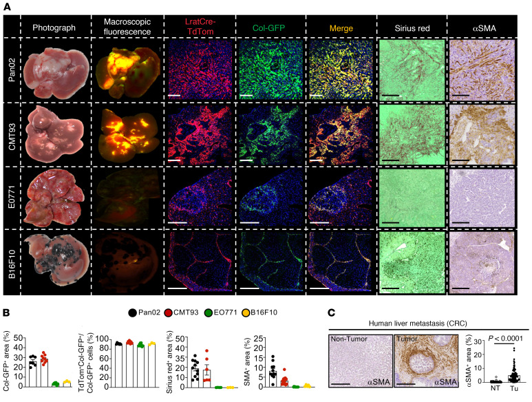
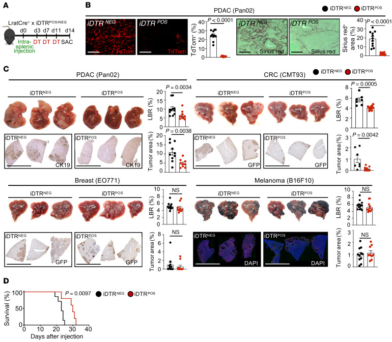
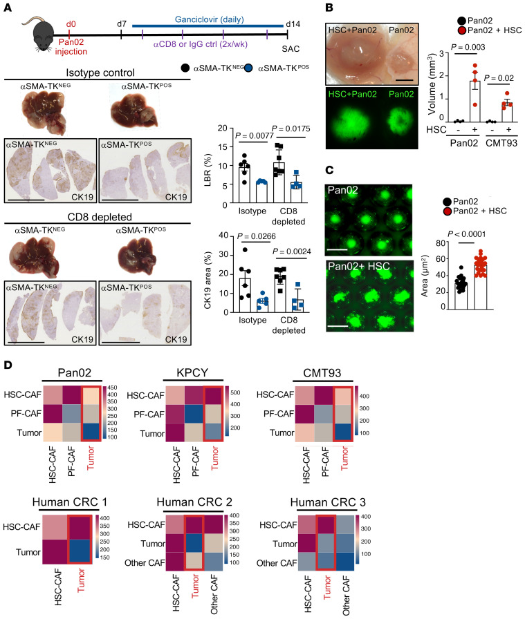
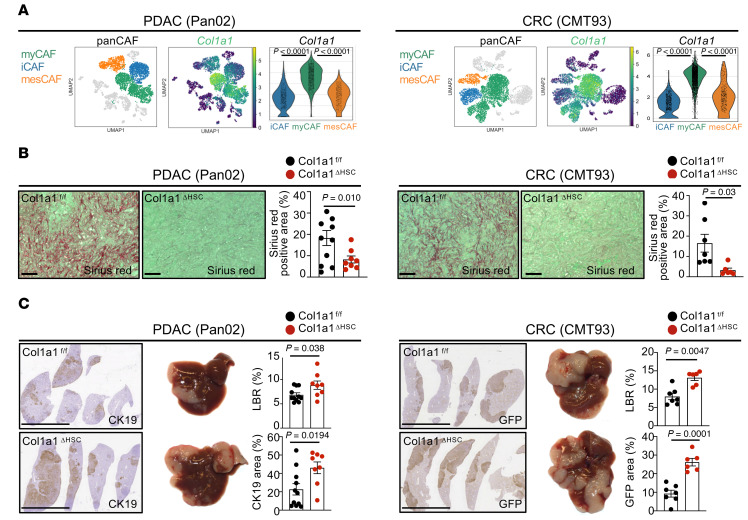
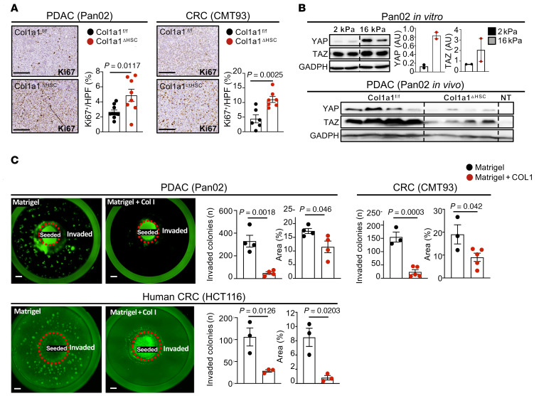
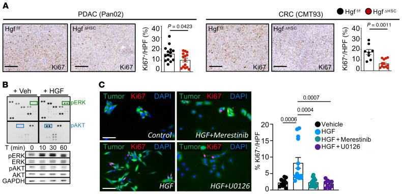
Cancer-associated fibroblasts (CAF) may exert tumor-promoting and tumor-suppressive functions, but the mechanisms underlying these opposing effects remain elusive. Here, we sought to understand these potentially opposing functions by interrogating functional relationships among CAF subtypes, their mediators, desmoplasia, and tumor growth in a wide range of tumor types metastasizing to the liver, the most common organ site for metastasis. Depletion of hepatic stellate cells (HSC), which represented the main source of CAF in mice and patients in our study, or depletion of all CAF decreased tumor growth and mortality in desmoplastic colorectal and pancreatic metastasis but not in nondesmoplastic metastatic tumors. Single-cell RNA-Seq in conjunction with CellPhoneDB ligand-receptor analysis, as well as studies in immune cell-depleted and HSC-selective knockout mice, uncovered direct CAF-tumor interactions as a tumor-promoting mechanism, mediated by myofibroblastic CAF-secreted (myCAF-secreted) hyaluronan and inflammatory CAF-secreted (iCAF-secreted) HGF. These effects were opposed by myCAF-expressed type I collagen, which suppressed tumor growth by mechanically restraining tumor spread, overriding its own stiffness-induced mechanosignals. In summary, mechanical restriction by type I collagen opposes the overall tumor-promoting effects of CAF, thus providing a mechanistic explanation for their dual functions in cancer. Therapeutic targeting of tumor-promoting CAF mediators while preserving type I collagen may convert CAF from tumor promoting to tumor restricting.

Keywords: Cancer; Collagens; Fibrosis; Hepatology; Oncology.


Conflict of interest statement

Conflict of interest: The authors have declared that no conflict of interest exists.

Figures

Figure 1
Figure 1. CAF ontogeny in mouse and human liver metastasis.
(A) Tumor images and their macroscopic fluorescence of LratCre-TdTom and collagen1a1-GFP; confocal microscopy of LratCre-TdTom, collagen1a1-GFP (Col-GFP), Sirius red, and αSMA IHC in Pan02, CMT93, EO771, and B16F10 liver metastasis. Scale bars: 100 μm. (B) Quantification of data in A. Data are shown as the mean ± SEM; n = 3–9 mice/cell line. (C) Representative IHC and quantifications of αSMA in human CRC liver metastasis (n = 100). NT, nontumor; Tu, tumor. Scale bars: 100 μm. Data are shown as the mean ± SEM; paired t test.
Figure 2
Figure 2. CAF subpopulations in mouse and human liver metastasis.
(A) UMAPs of scRNA-Seq of murine PDAC and CRC liver metastasis (n = 1 each) and human CRC metastasis (n = 6), displaying HSC and PF as a percentage of panCAF; murine HSC markers Lrat, Lum, and Pdgfrb; murine PF marker Msln, Upk1b, and Upk3b; human HSC markers PDGFRB, GEM, and FRZB; and human PF markers MSLN, UPK1B, and UPK3B. (B) UMAPs showing percentages and markers of myCAF, iCAF, and mesCAF subpopulations for murine PDAC models Pan02 and KPCY (n = 1 each), murine CRC model CMT93 (n = 1) and human CRC liver metastasis (n = 6).
Figure 3
Figure 3. HSC-derived CAF promote desmoplastic tumor growth and mortality.
(A and B) HSC-CAF depletion via DT injection (0.5 ng/g BW) in LratCre+TdTom+iDTR+ or LratCre+TdTom+iDTR littermates (B) reduces TdTom expression and Sirius red in tumors. Scale bars: 100 μm. (C) Representative macroscopic and IHC images of liver and liver-to-body weight ratio (LBR) and tumor area quantifications (n = 7–15 mice/group), showing effects of HSC-CAF depletion on desmoplastic (Pan02, CMT93) but not on nondesmoplastic (EO771, B16F10) liver metastasis. Scale bars: 1 cm. (D) Survival studies in iDTR mice injected with DT until death (n = 5–8 mice/group for each model). Statistics were done by 2-tailed unpaired t test or Mann Whitney U (B and C) or Mantel-Cox test (D). Data are shown as the mean ± SEM.
Figure 4
Figure 4. αSMA+ CAF promote desmoplastic tumor growth and mortality.
(A–C) CAF depletion in αSMA-TK mice, injected daily from day 3 after surgery with ganciclovir (10 mg/kg) (B) reduces αSMA IHC (n = 9–10 mice/group) and Sirius red (n = 9–13 mice/group). Scale bars: 100 μm. (C) Representative macroscopic and IHC images of liver and liver-to-body weight ratio (LBR) and tumor area quantification, showing the effects of CAF depletion on Pan02 and CMT93 liver metastasis (n = 5–13 mice/group). Scale bars: 1 cm. (D) Survival studies in αSMA-TK mice injected with ganciclovir until death (n = 5–8 mice/group for each model). Statistics were done by 2-tailed unpaired t test or Mann Whitney U (B and C) or Mantel-Cox test (D). Data are shown as the mean ± SEM.
Figure 5
Figure 5. CAF depletion in advanced stages arrests growth and promotes survival.
(A) HSC-CAF depletion via DT injection (0.5 ng/g) in LratCre+TdTom+iDTR+ or LratCre+TdTom+iDTR littermates. (B and C) Representative macroscopic and IHC images of liver and liver-to-body weight ratio (LBR) and tumor area quantification (n = 6–8 mice/group), showing the effects of late HSC-CAF depletion on desmoplastic tumors (Pan02, CMT93). Scale bars: 1 cm. (D) Survival studies in iDTR mice injected with DT (n = 5 mice/group). DT was injected from day 13 onward at intervals of 3 days until the end of the study. (E) Contrast-CT scan analysis and tumor volume quantification of animals undergoing late HSC-CAF depletion via LratCre × iDTR. Arrows indicate metastatic tumors (dark areas); lighter areas show nontumor tissue. Statistics were done by 2-tailed unpaired t test or Mann Whitney U dependent on data distribution (B, C, and E) or Mantel-Cox test (D). Data are shown as the mean ± SEM.
Figure 6
Figure 6. CAF promote tumor cell proliferation through direct interaction.
(A) Representative IHC images and quantification of Ki67+ cells in Pan02 and CMT93 models in iDTR models of CAF depletion. Scale bars: 1 cm. (B) UMAPs of scRNA-Seq displaying all cell populations in Pan02, CMT93, and KPCY models and human CRC liver metastasis, with accompanying CellphoneDB analysis showing ligand-receptor interactions between different cell populations. CellphoneDB analysis was performed on 3 CRC liver metastasis samples of 6 merged samples based on sufficient cell numbers for both CAF and tumor cells. Statistics were performed using 2-tailed unpaired t test or Mann Whitney U test dependent on data distribution (A). Data are shown as the mean ± SEM.
Figure 7
Figure 7. CAF promote tumor growth through direct interaction.
(A) CD8+ T cell depletion or isotype injection in αSMA-TK mice injected with ganciclovir (i.p. 10 mg/kg) to deplete CAF simultaneously. Pan02 liver metastasis in αSMA-TK mice with representative macroscopic and IHC images of liver and liver-to-body weight ratio (LBR) and tumor area quantification (n = 5–7 mice/group). Scale bars: 1 cm. (B) Subcutaneous coinjection of HSC with tumor cells (Pan02 or CMT93) or tumor cells alone in immune-deficient Rag2–/– mice. (C) Representative pictures and quantification of cocultured spheroids comprising Pan02 cells alone and Pan02 cells with HSC. (D) CellphoneDB analysis showing ligand-receptor interactions between tumor cells with HSC-CAF and PF-CAF and/or other CAF in mouse and human liver metastasis samples. Statistics were performed using 2-tailed unpaired t test or Mann Whitney U test dependent on data distribution (A–C). Data are shown as the mean ± SEM.
Figure 8
Figure 8. Type 1 collagen restricts tumor growth in vivo.
(A) UMAPs and gene signatures of myCAF and Col1a1 in Pan02 and CMT93 (n = 2). (B and C) Col1a1fl/fl and Col1a1ΔHSC (n = 6–11 mice per group) were intrasplenically injected with Pan02 or CMT93 cells. (B) Sirius red staining. Scale bars: 100 μm. (C) Representative macroscopic and IHC images of liver and liver-to-body weight ratio (LBR) and tumor area quantification. Scale bars: 1 cm. Statistics were done using 2-tailed unpaired t test or Mann Whitney U test dependent on data distribution. Data are shown as the mean ± SEM.
Figure 9
Figure 9. Type 1 collagen restricts tumor growth independent of stiffness.
(A) Col1a1fl/fl and Col1a1ΔHSC (n = 6–11 mice per group) were intrasplenically injected with Pan02 or CMT93 cells. Representative images and quantification of Ki67+ cells. Scale bars: 100 μm. (B) YAP and TAZ Western blot and quantification in cells plated on soft (2 kPa) or stiff (16 kPa) plates and Western blot from Col1a1fl/fl and Col1a1ΔHSC nontumor and tumor tissue (nontumor, NT, n = 1; tumor n = 4 mice per group) in the Pan02 liver metastasis model. (C) Representative images and quantification of an in vitro collagen-Matrigel assay, with cells seeded in the center and invading cells in the periphery in Matrigel alone or Matrigel and type 1 collagen (5 mg/ml) day 10 or day 12 (HCT116) after seeding (n = 3–4 wells per group). Statistics were done using 2-tailed unpaired t test or Mann Whitney U test dependent on data distribution (A–C). Data are shown as the mean ± SEM.
Figure 10
Figure 10. myCAF-enriched HA promotes tumor growth in vivo.
(A) UMAPs and gene signatures of myCAF and Has2 in Pan02 and CMT93 (n = 1 each). (B) Efficient reduction of HA shown by IHC in Has2ΔHSC mice compared with Has2fl/fl littermates (NL, normal liver; NT, nontumor; Tu, tumor). (C) Representative IHC and quantification of HA staining in human CRC liver metastasis with matched nontumor tissue (n = 100). Scale bars: 100 μm. Statistics were done using Paired t test. (D) Representative macroscopic and IHC images of liver and liver-to-body weight ratio (LBR) and tumor area quantifications (n = 7–8 per group each). Scale bars: 1 cm. Statistics were done using 2-tailed unpaired t test or Mann Whitney U test dependent on data distribution (B and D) and Wilcoxon’s matched-pairs signed-rank test (C). Data are shown as the mean ± SEM.
Figure 11
Figure 11. HA promotes tumor cell growth and invasiveness.
(A) Representative IHC images and quantification of Ki67+ cells in Pan02 and CMT93 models in Has2fl/fl and Has2ΔHSC animals (n = 7–8 per group). Scale bars: 100 μm. (B and C) Representative images and quantification of in vitro Matrigel assay, with cells seeded in the center and invading cells in the periphery in Matrigel alone, Matrigel and high-molecular-weight HA, Matrigel and HA and type 1 collagen, or Matrigel and type 1 collagen (HA and type I collagen: 2.5 mg/ml) day 6 after seeding (n = 3–4 per group each). (D) Coinjection of Has2fl/fl and Has2ΔHSC with tumor cells (Pan02 or CMT93) in RAG2–/– mice. (E and F) Representative images and quantification of cocultured spheroids (D) or 2D cocultures (E) and Ki67+ cells comprising Has2fl/fl and Has2ΔHSC with Pan02 tumor cells. Statistics were done using 2-tailed unpaired t test or Mann Whitney U test dependent on data distribution (A and D–F) or 1-way ANOVA using Fisher’s LSD test (B and C). Data are shown as the mean ± SEM.
Figure 12
Figure 12. iCAF-enriched Hgf promotes tumor growth in vivo.
(A) UMAPs and gene signatures of iCAF and Hgf in Pan02 and CMT93 (n = 2). (B) Representative macroscopic and IHC images of liver and liver-to-body weight ratio (LBR) and tumor area quantification (n = 10–15 per group each). Scale bars: 1 cm. Statistics were done using 2-tailed unpaired t test or Mann Whitney U test dependent on data distribution. Data are shown as the mean ± SEM.
Figure 13
Figure 13. Hgf promotes tumor cell proliferation through Met receptor.
(A) Representative IHC images and quantification of Ki67+ cells in Pan02 and CMT93 models in Hgffl/fl and HgfΔHSC animals (n = 10–15 per group). Scale bars: 100 μm. (B) Pan02 cells treated with recombinant mouse HGF (25 ng/ml) or vehicle for 10 minutes followed by a phospho-kinase array and Western blot for phosphorylated and total ERK1/2 and AKT. (C) Pan02 cells treated with HGF (25 ng/ml) or Merestinib (3 mM) or U0126 (10 mM) and followed by recombinant mouse HGF (25 ng/ml) or vehicle for 48 hours followed by Ki67 staining. Scale bars: 250 μm. Statistics were done using Mann Whitney U test or unpaired t test dependent on data distribution (A) or 1-way ANOVA with Kruskal Wallis post hoc test (C). Data are shown as the mean ± SEM.

References

    1. Chiang AC, Massague J. Molecular basis of metastasis. N Engl J Med. 2008;359(26):2814–2823. doi: 10.1056/NEJMra0805239. - DOI - PMC - PubMed
    1. Griscom JT, Wolf PS, eds. Liver Metastasis. StatPearls Publishing; 2021. - PubMed
    1. Lambert AW, et al. Emerging biological principles of metastasis. Cell. 2017;168(4):670–691. doi: 10.1016/j.cell.2016.11.037. - DOI - PMC - PubMed
    1. Nguyen DX, et al. Metastasis: from dissemination to organ-specific colonization. Nat Rev Cancer. 2009;9(4):274–284. - PubMed
    1. Mizrahi JD, et al. Pancreatic cancer. Lancet. 2020;395(10242):2008–2020. doi: 10.1016/S0140-6736(20)30974-0. - DOI - PubMed

Publication types

MeSH terms

Substances