. 2021 Apr 27;17(4):e1009561.
doi: 10.1371/journal.ppat.1009561. eCollection 2021 Apr.

Genetic and biological properties of H7N9 avian influenza viruses detected after application of the H7N9 poultry vaccine in China

Affiliations

Genetic and biological properties of H7N9 avian influenza viruses detected after application of the H7N9 poultry vaccine in China

Xin Yin et al. PLoS Pathog. .

Abstract

The H7N9 avian influenza virus (AIV) that emerged in China have caused five waves of human infection. Further human cases have been successfully prevented since September 2017 through the use of an H7N9 vaccine in poultry. However, the H7N9 AIV has not been eradicated from poultry in China, and its evolution remains largely unexplored. In this study, we isolated 19 H7N9 AIVs during surveillance and diagnosis from February 2018 to December 2019, and genetic analysis showed that these viruses have formed two different genotypes. Animal studies indicated that the H7N9 viruses are highly lethal to chicken, cause mild infection in ducks, but have distinct pathotypes in mice. The viruses bound to avian-type receptors with high affinity, but gradually lost their ability to bind to human-type receptors. Importantly, we found that H7N9 AIVs isolated in 2019 were antigenically different from the H7N9 vaccine strain that was used for H7N9 influenza control in poultry, and that replication of these viruses cannot, therefore, be completely prevented in vaccinated chickens. We further revealed that two amino acid mutations at positions 135 and 160 in the HA protein added two glycosylation sites and facilitated the escape of the H7N9 viruses from the vaccine-induced immunity. Our study provides important insights into H7N9 virus evolution and control.


Conflict of interest statement

The authors have declared that no competing interests exist.

Figures

Fig 1
Fig 1. Phylogenetic analyses and genotypes of H7N9 highly pathogenic avian influenza viruses.
The phylogenetic trees of the HA (A) and NA (B) genes were rooted to A/chicken/Rostock/45/1934 (H7N1) and A/chicken/Italy/22A/1998 (H5N9), respectively. The viruses sequenced in this study are shown in red in the phylogenetic trees. (C) Genotypes of H7N9 and H7N2 viruses and the hosts in which these genotypes were detected. The genotypes of the viruses isolated between February 2017 and January 2018 were reported previously [7]; the viruses isolated between February 2018 and December 2019 were analyzed in this study. The numbers of strains of each genotype are provided in parentheses.
Fig 2
Fig 2. Replication and virulence of H7N9 viruses in mice.
(A) Viral titers in organs of mice after inoculation with 106 EID50 of different viruses. Three mice from each group were killed on Day 3 p.i., and virus titers were determined in eggs. Color bars show the mean, and the error bars represent standard deviations. The values labeled with a red star indicate that the virus was only detected in the organ of one mouse. The dashed lines indicate the lower limit of virus detection. (B) Changes in body weight in the groups of five mice after inoculation with 106 EID50 of different viruses. (C) Mouse-lethal doses of the CK/IM/SD010/19 virus.
Fig 3
Fig 3. Receptor-binding properties of H7N9 representative viruses isolated between 2013 and 2019.
The binding of H7N9 viruses to two different glycans (α-2,3-glycans, blue; α-2,6-glycans, pink) was assessed. The data shown are the means of three repeats, the error bars indicate standard deviations.
Fig 4
Fig 4. Antigenic cartography of H7N9 viruses.
The antigenic map was generated by using the HI assay data shown in S2 Table. Each unit in the coordinate represents a 2-fold difference in HI titer. The pink cubes represent the antisera generated from the indicated viruses. The red balls indicate the viruses used for antisera generation, and the green balls show the test viruses.
Fig 5
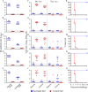
Fig 5. Protective efficacy of H5/H7-Re2 trivalent inactivated vaccine against challenge with different H7N9 viruses in chickens.
HI antibody titers (A-E), virus shedding titers (F-J), and survival patterns (K-O) of chickens challenged with the H7N9 highly pathogenic viruses CK/GX/SD098/17 (A, F, and K), CK/SX/SD006/18 (B, G, and L), PCK/LN/SD004/19 (C, H, and M), CK/IM/SD010/19 (D, I, and N), and CK/LN/SD25/19 (E, J, and O). The dashed lines shown in A-E show the cutoff value for seroconversion and those in F-J show the lower limit of virus detection. Virus titers shown in F-J are the means from the birds that survived. A value of 0.5 was assigned to virus shedding-negative birds for statistical purposes. The asterisks indicate that the bird(s) died before that day, and therefore virus shedding data were not available for statistical analysis. All of the chickens in these control groups died within 5 days of challenge. The letter “a” indicates p < 0.001 compared with the corresponding titers of the control birds.
Fig 6
Fig 6. Key mutations in HA that contributed to the antigenic drift of the 2019 H7N9 viruses.
(A) Amino acid differences in the HA1 protein of the representative H7N9 viruses CK/GX/SD098/17 and CK/IM/SD010/19. The key amino acids in the head of the HA1 trimer that differ between the two viruses are shown in red. The colored boxes show different antigenic regions (site A to site E). The 2D structure of the HA1 protein of CK/GX/SD098/17 (B) and CK/IM/SD010/19 (C), and the 3D structure of the HA1 protein of CK/GX/SD098/17 (D) were obtained by using SWISS-MODEL; images were drawn with Pymol software. The numbers show the positions of the key amino acid in the head of the HA1 trimer that are different in the representative viruses. (E) HI titers of different H7N9 mutants against H7-Re2 antiserum and H7N9 monoclonal antibodies. (F) Mobility of H7N9 avian influenza HA1 protein analyzed by SDS-PAGE and Western blotting.

References

    1. WHO/GIP. Monthly Risk Assessment Summary. World Health Organization. 2020. https://www.who.int/influenza/human_animal_interface/Influenza_Summary_I....
    1. Zhang Q, Shi J, Deng G, Guo J, Zeng X, He X, et al.. H7N9 Influenza Viruses Are Transmissible in Ferrets by Respiratory Droplet. Science. 2013;341(6144):410–4. 10.1126/science.1240532 WOS:000322259200053. - DOI - PubMed
    1. Pantin-Jackwood MJ, Miller PJ, Spackman E, Swayne DE, Susta L, Costa-Hurtado M, et al.. Role of poultry in the spread of novel H7N9 influenza virus in China. J Virol. 2014;88(10):5381–90. Epub 2014/02/28. 10.1128/JVI.03689-13 - DOI - PMC - PubMed
    1. Kalthoff D, Bogs J, Grund C, Tauscher K, Teifke JP, Starick E, et al.. Avian influenza H7N9/13 and H7N7/13: a comparative virulence study in chickens, pigeons, and ferrets. J Virol. 2014;88(16):9153–65. Epub 2014/06/06. 10.1128/JVI.01241-14 - DOI - PMC - PubMed
    1. Yang L, Zhu W, Li X, Chen M, Wu J, Yu P, et al.. Genesis and Spread of Newly Emerged Highly Pathogenic H7N9 Avian Viruses in Mainland China. J Virol. 2017;91(23). Epub 2017/09/29. 10.1128/JVI.01277-17 - DOI - PMC - PubMed

Publication types

MeSH terms

Substances