. 2021 May 4;35(5):109055.
doi: 10.1016/j.celrep.2021.109055. Epub 2021 Apr 13.

SARS-CoV-2 infection of primary human lung epithelium for COVID-19 modeling and drug discovery

Affiliations

SARS-CoV-2 infection of primary human lung epithelium for COVID-19 modeling and drug discovery

Apoorva Mulay et al. Cell Rep. .

Abstract

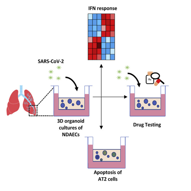
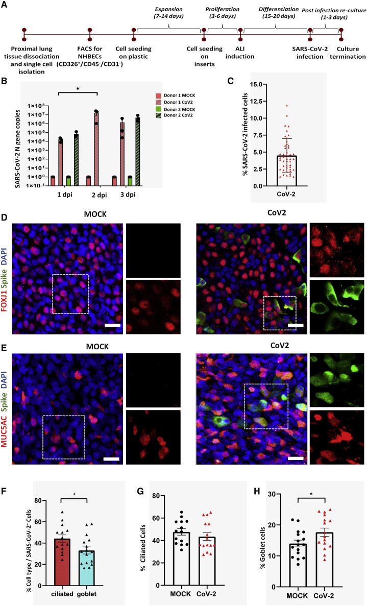
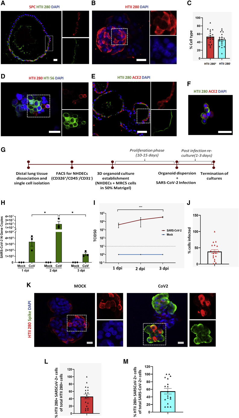
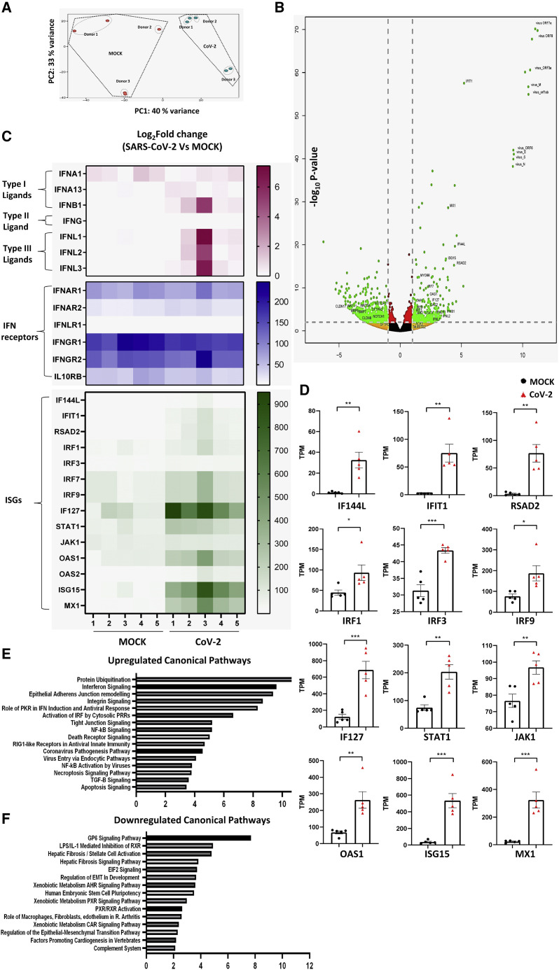
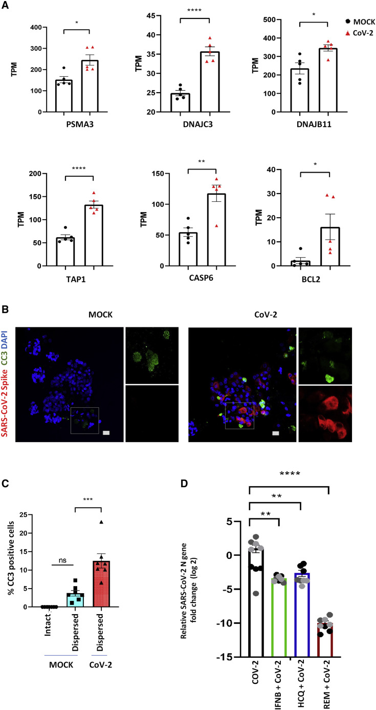
Coronavirus disease 2019 (COVID-19) is the latest respiratory pandemic caused by severe acute respiratory syndrome-related coronavirus 2 (SARS-CoV-2). Although infection initiates in the proximal airways, severe and sometimes fatal symptoms of the disease are caused by infection of the alveolar type 2 (AT2) cells of the distal lung and associated inflammation. In this study, we develop primary human lung epithelial infection models to understand initial responses of proximal and distal lung epithelium to SARS-CoV-2 infection. Differentiated air-liquid interface (ALI) cultures of proximal airway epithelium and alveosphere cultures of distal lung AT2 cells are readily infected by SARS-CoV-2, leading to an epithelial cell-autonomous proinflammatory response with increased expression of interferon signaling genes. Studies to validate the efficacy of selected candidate COVID-19 drugs confirm that remdesivir strongly suppresses viral infection/replication. We provide a relevant platform for study of COVID-19 pathobiology and for rapid drug screening against SARS-CoV-2 and emergent respiratory pathogens.

Keywords: COVID-19; SARS-CoV-2; adult lung epithelium; alveolar type 2 cells; alveoli; drug discovery; interferon; primary in vitro model; remdesivir.


Conflict of interest statement

Declaration of interests The authors declare no competing interests.

Figures

None
Graphical abstract
Figure 1
Figure 1
SARS-CoV-2 infects normal human proximal airway cells (A) Workflow for establishment of human proximal airway ALI cultures and their infection with SARS-CoV-2. (B) ALI cultures of proximal airway epithelial cells are susceptible to SARS-CoV-2 infection, indicated by viral N gene copy numbers, which peaked at 2 days postinfection (dpi); n = 3–6 cultures from 2 independent donors. Red and green colors indicate cultures from separate donors. Data were analyzed using one-way ANOVA with Tukey’s post hoc correction and represented N gene copy numbers for individual cultures ± SEM. p < 0.05. (C) Percentage of cells from proximal airway ALI cultures infected with SARS-CoV-2 at 2 dpi. n = 14–15 fields from two biological replicates. (D and E) FOXJ1-positive ciliated cells (red) (D) and MUC5AC-positive goblet cells (red) (E) infected by SARS-CoV-2 (green). Scale bar, 20 μm. Image magnification = 2.3× with a 20× objective. (F) Percentage of infected cells that are either ciliated cells or goblet cells. (G and H) Change in the percentage of (G) ciliated and (H) goblet cells after SARS-CoV-2 infection. (F–H) n = 7–8 fields from two biological replicates. Data are presented as mean ± SEM (significance is determined by two-tailed t test). p < 0.05.
Figure 2
Figure 2
SARS-CoV-2 infects normal human distal AT2 cells (A) Sections of Matrigel-embedded alveospheres showing colocalization of AT2 markers HTII-280 and SPC. (B) Whole-mount staining of alveospheres dispersed from Matrigel for AT2 marker HTII-280. Scale bar = 20 μm. (C) Percentage of HTII-280-positive and HTII-280-negative cells in alveospheres in n = 3–5 fields from three biological replicates (two-tailed t test). (D) Whole-mount alveospheres stained for the AT2 marker HTII-280 and AT1 cell marker HTI 56. (E and F) ACE2 staining in sectioned alveospheres (E) and in AT2 cells (F) dissociated from alveospheres. Scale bar = 20 μm. (G) Workflow for establishment of human distal alveolar cultures and their infection with SARS-CoV-2. (H) Absolute N gene copy numbers in SARS-CoV-2-infected cultures peaked at 2 dpi; n = 3 independent cultures. Data were analyzed using two-way ANOVA with Sidak’s post hoc correction and represented as N gene copy number for individual cultures ± SEM. ∗p <0.05. (I) Viral load in supernatant increased from 1 to 3 dpi. ∗∗∗p < 0.001. (J) Percentage of total cells infected (n = 3). (K) Infection was assessed at 2 dpi by antibody against SARS-CoV-2 “spike” protein (green) and colocalized with viral AT2 cell marker HTII-280 (red). Scale bar, 20 μm. (L) Percentage of HTII-280-positive cells infected by SARS-CoV-2; n = 3–5 fields from each of three biological replicates. (M) Percentage of total infected cells that are HTII-280-positive AT2 cells; n = 3–5 fields from each of three biological replicates.
Figure 3
Figure 3
Primary human alveolar cultures as a model to study SARS-CoV-2-induced host response (A) Principal-component analysis of infected alveolar cultures and MOCK cultures showing variance in the global transcriptome of the two groups (n = 5 from three biological replicates for both MOCK and SARS-CoV-2-infected cultures). (B) Volcano plot of gene expression changes in SARS-CoV-2-infected versus mock cultures defined by p value and >2-fold change. (C) Heatmap of normalized TPM counts for various interferon ligands, receptors, and ISGs. (D) Quantification of normalized TPM counts of various downstream ISGs between MOCK and SARS-CoV-2-infected cultures (n = 5; significance determined by two-tailed t test). p < 0.05, ∗∗p < 0.01, ∗∗∗p < 0.001, ∗∗∗∗p < 0.0001. (E and F) The most highly upregulated (E) and downregulated (F) canonical pathways in SARS-CoV-2-infected alveolar cultures.
Figure 4
Figure 4
SARS-CoV-2 infection triggers apoptosis of alveolar cells (A) Quantification of normalized TPM counts for apoptosis-related genes between MOCK- and SARS-CoV-2 infected alveolar cultures (n = 5; significance is determined by two-tailed t test). p < 0.05, ∗∗p < 0.01, ∗∗∗p < 0.001, ∗∗∗∗p < 0.0001. (B) Infected AT2 cells (red) also demonstrated signs of cellular apoptosis at 3 dpi, indicated by positive staining for cleaved caspase-3 (green). Scale bar: 20 μm. (C) Comparison of percentage of cells staining positive for CC3 in intact Matrigel-embedded MOCK alveospheres, alveolar cultures dispersed from Matrigel, and infected cultures dispersed from Matrigel. n = 7 (one-way ANOVA with Tukey’s post hoc test). p < 0.05, ∗∗p < 0.01, ∗∗∗p < 0.001, ∗∗∗∗p < 0.0001. (D) Pre-treatment of alveolar cultures with IFNB1, hydroxychloroquine, and remdesivir significantly reduced viral replication. The effect of remdesivir on viral replication was more pronounced than that of IFNB1 or hydroxychloroquine. Data are represented as log2 fold change for individual cultures ± SEM, normalized to mean infection and analyzed using one-way ANOVA with Tukey’s post hoc test. ∗∗p = 0.0012 for IFNB1, ∗∗p = 0.0044 for HCQ, and ∗∗∗∗p < 0.0001 for remdesivir. The three different colors indicate cultures from different biological replicates.

Update of

References

    1. Barkauskas C.E., Cronce M.J., Rackley C.R., Bowie E.J., Keene D.R., Stripp B.R., Randell S.H., Noble P.W., Hogan B.L. Type 2 alveolar cells are stem cells in adult lung. J. Clin. Invest. 2013;123:3025–3036. - PMC - PubMed
    1. Beigel J.H., Tomashek K.M., Dodd L.E., Mehta A.K., Zingman B.S., Kalil A.C., Hohmann E., Chu H.Y., Luetkemeyer A., Kline S. Remdesivir for the Treatment of Covid-19—Preliminary Report. N. Engl. J. Med. 2020;383:1813–1826. - PMC - PubMed
    1. Blanco-Melo D., Nilsson-Payant B.E., Liu W.C., Uhl S., Hoagland D., Møller R., Jordan T.X., Oishi K., Panis M., Sachs D. Imbalanced Host Response to SARS-CoV-2 Drives Development of COVID-19. Cell. 2020;181:1036–1045.e9. - PMC - PubMed
    1. Cagno V. SARS-CoV-2 cellular tropism. Lancet Microbe. 2020;1:e2–e3. - PMC - PubMed
    1. Carraro G., Mulay A., Yao C., Mizuno T., Konda B., Petrov M., Lafkas D., Arron J.R., Hogaboam C.M., Chen P. Single-Cell Reconstruction of Human Basal Cell Diversity in Normal and Idiopathic Pulmonary Fibrosis Lung. Am. J. Respir. Crit. Care Med. 2020;202:1540–1550. - PMC - PubMed

Publication types

MeSH terms