. 2021 Jun:273:120806.
doi: 10.1016/j.biomaterials.2021.120806. Epub 2021 Apr 9.

Effect of tissue microenvironment on fibrous capsule formation to biomaterial-coated implants

Affiliations

Effect of tissue microenvironment on fibrous capsule formation to biomaterial-coated implants

Jamie L Hernandez et al. Biomaterials. 2021 Jun.

Abstract

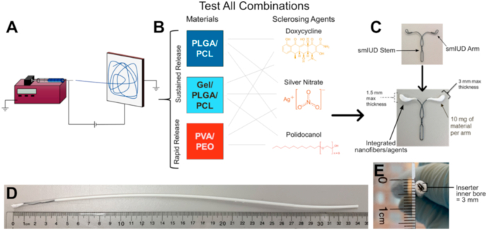
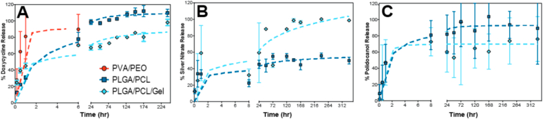
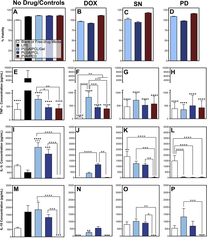
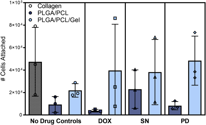
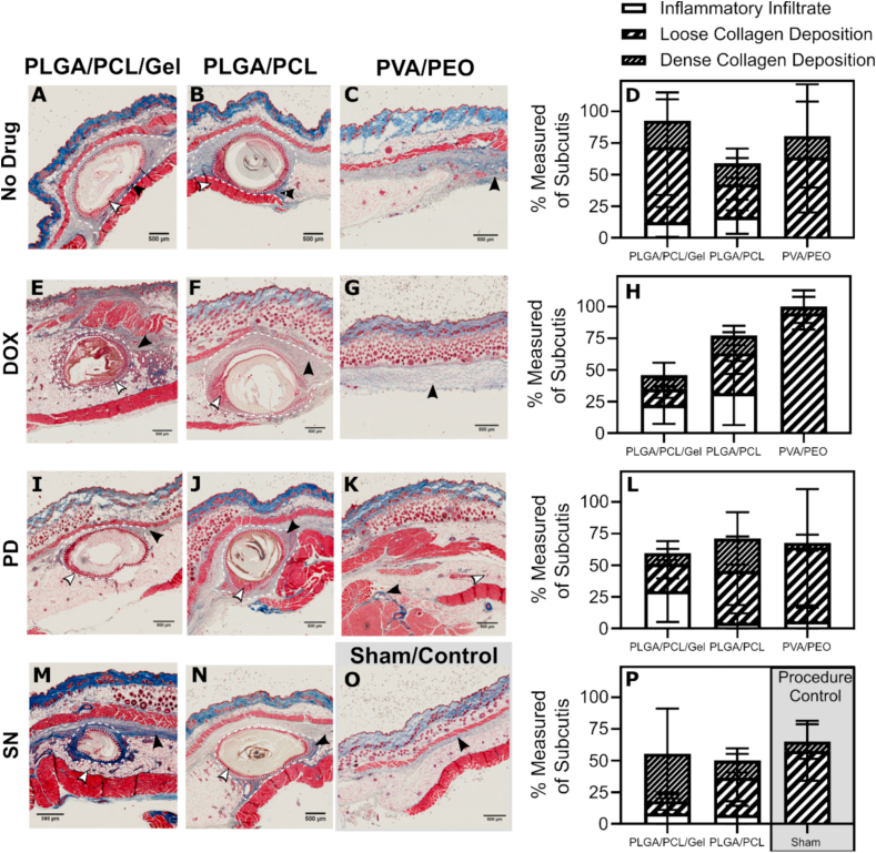
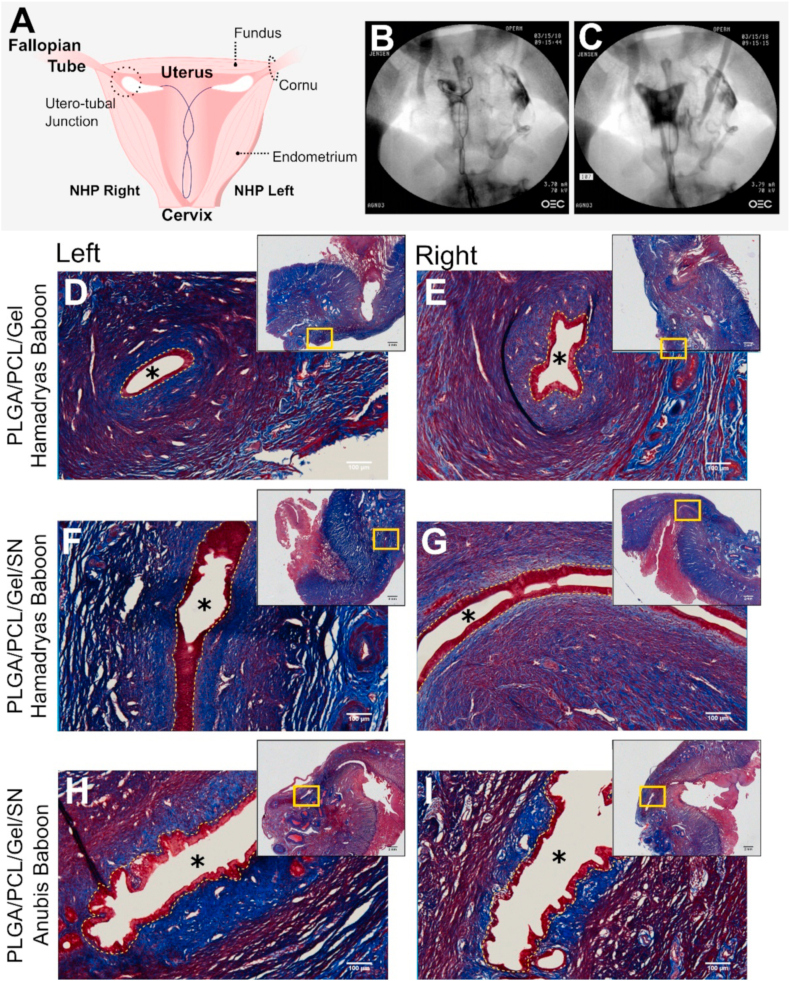
Within tissue exposed to the systemic immune system, lymphocytes and fibroblasts act against biomaterials via the development of a fibrous capsule, known as the foreign body reaction (FBR). Inspired by the natural tolerance that the uterine cavity has to foreign bodies, our study explores the role of microenvironment across classical (subcutaneous) and immune privileged (uterine) tissues in the development of the FBR. As a model biomaterial, we used electrospun fibers loaded with sclerosing agents to provoke scar tissue growth. Additionally, we integrated these materials onto an intrauterine device as a platform for intrauterine biomaterial studies. Polyester materials in vitro achieved drug release up to 10 days, greater pro-inflammatory and pro-healing cytokine expression, and the addition of gelatin enabled greater fibroblast attachment. We observed the materials that induced the greatest FBR in the mouse, had no effect when inserted at the utero-tubal junction of non-human primates. These results suggest that the FBR varies across different tissue microenvironments, and a dampened fibrotic response exists in the uterine cavity, possibly due to immune privilege. Further study of immune privileged tissue factors on biomaterials could broaden our understanding of the FBR and inform new methods for achieving biocompatibility in vivo.

Keywords: Electrospun fibers; Female reproductive tract; Foreign body reaction; Immune privilege; Intrauterine device; Sclerosing agents.


Conflict of interest statement

The authors declare the following financial interests/personal relationships which may be considered as potential competing interests: Dr. Jensen has received payments for consulting from Abbvie, Cooper Surgical, Bayer Healthcare, Evofem, Mayne Pharma, Merck, Sebela, and TherapeuticsMD. OHSU has received research support from Abbvie, Bayer Healthcare, Daré, Estetra SPRL, Medicines360, Merck, and Sebela. These companies and organizations may have a commercial or financial interest in the results of this research and technology. These potential conflicts of interest have been reviewed and managed by OHSU. Bob Katz is a paid consultant for Sebela Pharmaceuticals and CEO of Hybridge Medical, LLC.

Figures

Image 1
Graphical abstract
Fig. 1
Fig. 1
Material design and integration onto an IUD. (A) Schematic of the electrospinning process, and (B) the material drug combinations tested throughout this study. (C) Image of the modified VeraCept® nitinol wire IUD frame (top) and the proposed combined IUD/fiber device with required dimensions for the wrapped electrospun fibers (bottom). Fiber dimensions were constrained by the IUD insertion tube required for placement of the (D) folded fiber-IUD. The (E) inner bore of the inserter is 3 mm in diameter.
Fig. 2
Fig. 2
Different fiber formulations vary the release rate of sclerosing agents. Release of (A) Dox, (B) SN, and (C) PD from all fiber blends as percent of total theoretical loading plotted over time from n = 3 fiber sections (mean ± standard deviation).
Fig. 3
Fig. 3
Fibers/drug formulations affect the expression of pro- and anti-inflammatory cytokines by macrophages. (A–D) Cell viability and expression of proinflammatory cytokines (E–H) TNF-α,(I–L) IL-1β, and anti-inflammatory cytokine (M − P) IL-10 from RAW 264.7 macrophages 48-h post-treatment (mean + standard deviation). Viability results are measured for n = 1 cultured fiber sections and n = 3 assay replicates as a percent signal against untreated cells. Cytokine results are measured from n = 3 fiber sections, n = 3 assay replicates. Statistically significant different expression than media (◇) and LPS (◆) is marked on all plots. Statistical differences between material types (*) are specifically marked. For each symbol, * = p ≤ 0.05, ** = p ≤ 0.01, *** = p ≤ 0.001, and **** = p ≤ 0.0001.
Fig. 4
Fig. 4
Fiber formulations contribute significantly to fibroblast attachment. Plot of the cell number att atached on the fibers after 24 h as measured by a standard MTS assay. Collagen treated coverslips were used as an attachment positive control. Cell number attached was measured on n = 3 cultured material segments and is plotted as individual measurements with the mean ± standard deviation.
Fig. 5
Fig. 5
Fiber formulations induce varying degrees of fibrosis: Masson's Trichrome stained tissue sections for (A–C) fiber implants loaded (A–C) without drug, (E–G) doxycycline, (I–K) polidocanol, (M − N) silver nitrate, and (O) the sham/procedure control. Implants are composed of (A, E, I, and L) PLGA/PCL/Gel, (B, F, J, and N) PLGA/PCL, or (C, G, and K) PVA/PEO. Collagen deposition is visualized in blue, cell cytoplasm is stained pink/red. Regions of inflammation (white arrows) and collagen deposition (black arrows) are marked. Images were taken at 2.5× magnification. Quantitative analysis of the histology images is included for fibers with (D) no drug, (H) Dox, (L) PD, and (P) SN/sham. Implant studies and analysis were conducted with n = 1 mouse and n = 2 implant pockets, with the exception of PLGA/PCL/PD and PLGA/PCL/Gel/Dox implants, which were analyzed using two images from a single implant replicate. Percent inflammatory infiltrate and total collagen deposition (loose and dense staining) of the subcutis were determined from representative implant images for each implant (n = 3 sections). Measurements are plotted as mean % ± standard deviation.
Fig. 6
Fig. 6
Fibers can be integrated onto IUD devices for intrauterine placement in a non-human primate model. (A) Diagram of the fiber-IUD and relevant anatomical features of the female reproductive tract. Fluoroscopy images of fiber loaded IUD placed in an anubis baboon with active fibers (B) without and (C) with contrast to outline shape of uterine cavity. Resulting tissue sections of left and right cornu of the uterus stained with Masson's trichrome 28-days following treatment with (D&E) PLGA/PCL/Gel fibers without drug in an hamadryas baboon (n = 1), (F & G) 60% (w/w) SN loaded fibers in the anubis baboon (n = 1), and (H&I) SN loaded fibers treated in the hamadryas baboon (n = 1). Main images are captured at 10× magnification, and overview scan images of the tissue sections are included at the top right. Asterisks (*) mark the lumen of the fallopian tube, and yellow-dashed lines outline the tubal epithelium.

Similar articles

Cited by

References

    1. Klopfleisch R., Jung F. The pathology of the foreign body reaction against biomaterials. J. Biomed. Mater. Res. 2017;105:927–940. - PubMed
    1. Klopfleisch R. Macrophage reaction against biomaterials in the mouse model – phenotypes, functions and markers. Acta Biomater. 2016;43:3–13. - PubMed
    1. Bryers J.D., Giachelli C.M., Ratner B.D. Engineering biomaterials to integrate and heal: the biocompatibility paradigm shifts. Biotechnol. Bioeng. 2012;109:1898–1911. - PMC - PubMed
    1. Anderson J.M., Rodriguez A., Chang D.T. Foreign body reaction to biomaterials. Semin. Immunol. 2008;20:86–100. - PMC - PubMed
    1. Mariani E., Lisignoli G., Borzì R.M., Pulsatelli L. Biomaterials: foreign bodies or tuners for the immune response? Int. J. Mol. Sci. 2019;20:1–42. - PMC - PubMed

Publication types

Substances