. 2021 Jul 13;144(2):144-158.
doi: 10.1161/CIRCULATIONAHA.120.052788. Epub 2021 Apr 28.

Salt Transiently Inhibits Mitochondrial Energetics in Mononuclear Phagocytes

Affiliations

Salt Transiently Inhibits Mitochondrial Energetics in Mononuclear Phagocytes

Sabrina Geisberger et al. Circulation. .

Erratum in

Abstract

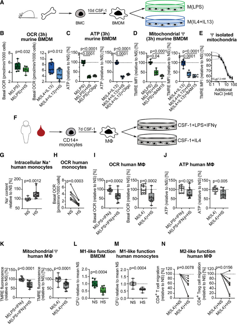
Background: Dietary high salt (HS) is a leading risk factor for mortality and morbidity. Serum sodium transiently increases postprandially but can also accumulate at sites of inflammation affecting differentiation and function of innate and adaptive immune cells. Here, we focus on how changes in extracellular sodium, mimicking alterations in the circulation and tissues, affect the early metabolic, transcriptional, and functional adaption of human and murine mononuclear phagocytes.

Methods: Using Seahorse technology, pulsed stable isotope-resolved metabolomics, and enzyme activity assays, we characterize the central carbon metabolism and mitochondrial function of human and murine mononuclear phagocytes under HS in vitro. HS as well as pharmacological uncoupling of the electron transport chain under normal salt is used to analyze mitochondrial function on immune cell activation and function (as determined by Escherichiacoli killing and CD4+ T cell migration capacity). In 2 independent clinical studies, we analyze the effect of a HS diet during 2 weeks (URL: http://www.clinicaltrials.gov. Unique identifier: NCT02509962) and short-term salt challenge by a single meal (URL: http://www.clinicaltrials.gov. Unique identifier: NCT04175249) on mitochondrial function of human monocytes in vivo.

Results: Extracellular sodium was taken up into the intracellular compartment, followed by the inhibition of mitochondrial respiration in murine and human macrophages. Mechanistically, HS reduces mitochondrial membrane potential, electron transport chain complex II activity, oxygen consumption, and ATP production independently of the polarization status of macrophages. Subsequently, cell activation is altered with improved bactericidal function in HS-treated M1-like macrophages and diminished CD4+ T cell migration in HS-treated M2-like macrophages. Pharmacological uncoupling of the electron transport chain under normal salt phenocopies HS-induced transcriptional changes and bactericidal function of human and murine mononuclear phagocytes. Clinically, also in vivo, rise in plasma sodium concentration within the physiological range reversibly reduces mitochondrial function in human monocytes. In both a 14-day and single meal HS challenge, healthy volunteers displayed a plasma sodium increase of [Formula: see text] and [Formula: see text] respectively, that correlated with decreased monocytic mitochondrial oxygen consumption.

Conclusions: Our data identify the disturbance of mitochondrial respiration as the initial step by which HS mechanistically influences immune cell function. Although these functional changes might help to resolve bacterial infections, a shift toward proinflammation could accelerate inflammatory cardiovascular disease.

Keywords: bacterial killing, humans; complex II; macrophages; metabolism; mitochondrial respiration; monocytes; salt.


Figures

Figure 1.
Figure 1.
High salt inhibits mitochondrial respiration in murine and human mononuclear phagocytes. A, Pictogram of the generation and activation of murine bone marrow–derived macrophages (BMDMs). B through D, BMDMs were treated for 3 hours with lipopolysaccharide (LPS) or interleukin (IL) 4+IL13, under normal salt (NS) or high salt (HS) conditions. (B) Basal oxygen consumption rate (OCR) (pooled data n=10 from 3 independent experiments). Data are depicted as box and whisker with minimum to maximum. Significance was analyzed by unpaired, 2-tailed t test. C, Relative ATP content, with Oligomycin A (Oligo, 10 µM) treatment as positive control for ATP decrease (pooled data n=12 from 2 independent experiments). Data are depicted as box and whisker with minimum to maximum. Significance was analyzed by 1-way ANOVA with Tukey’s post hoc test. D, Relative TMRE mean fluorescence intensity (MFI), with mitochondrial uncoupler BAM15 (10 µM) as positive control for mitochondrial depolarization (pooled data n=10 from 2 independent experiments). Data are depicted as box and whisker with minimum to maximum. Significance was analyzed by Kruskal-Wallis test with Dunn’s post hoc test (LPS groups) or by one-way ANOVA with Tukey’s post hoc test (IL4+IL13 groups). E, Relative TMRE MFI in isolated unactivated BMDM mitochondria, treated with increasing concentrations of NaCl (serial dilutions from 64 mM to 1 mM NaCl) for 3 hours (n=4). F, Pictogram of the generation and activation of human monocyte-derived macrophages. G, Relative intracellular Na+ content in human monocytes after 1 hour of HS treatment (n=13). Significance was analyzed by unpaired, two-tailed t test. H, Basal OCR in freshly isolated human monocytes (n=8 male donors), treated for 3 hours with 40 mM of NaCl (HS). Data are shown as donor-paired mean and were analyzed by paired, 2-tailed t test. I through K, Human monocyte-derived macrophages (MΦ) activated for 6 hours with LPS+ interferon (IFN) γ or IL4, under NS or HS conditions. I, Basal OCR (n=14 for M[LPS+INFγ] NS and HS, n=15 for M[IL4] NS, and n=12 for M[IL4] HS). J, ATP level in relation to NS (n=11 for NS, n=12 for HS for both M[LPS+INFγ] and M[IL4], respectively). K, TMRE fluorescence intensity in relation to NS (n=14 for M[LPS+INFγ] NS and HS, n=15 for M[IL4] NS and HS). I through K, Data are shown as box and whisker with minimum to maximum and were analyzed by unpaired, 2-tailed t test. Relative E. coli colony-forming units in murine BMDMs (L) (pooled data n=9 from 3 independent experiments) and human monocytes (M) (n=8), treated under NS or HS conditions, infected with E. coli for 3 hours. Data are shown as box and whisker with minimum to maximum in relation to NS and were analyzed by unpaired, 2-tailed t test. N, Effect of human M2 macrophages treated with NS and HS on CD4+ T cell and CD4+ regulatory T cell (Treg) migration (n=8 each). Data are shown as donor-paired mean and were analyzed by Wilcoxon matched-pairs signed-rank 2-tailed test. ψ indicates mitochondrial membrane potential; CFU, colony-forming unit; HS, high salt; NS, normal salt; and TMRE, tetramethylrhodamine ethyl ester.
Figure 2.
Figure 2.
High salt inhibits mitochondrial electron transport chain at complex II. A, Pictogram of the central carbon metabolism and ETC. Glucose-derived 13C-label is represented as red dots. Glutamine-derived 13C-label is represented as blue dots. Mitochondrial membrane potential is ψ. B through D, Bone marrow–derived macrophages (BMDMs) treated for 3 h (B and D) or 24 h (C) with lipopolysaccharide (LPS) or interleukin (IL) 4+IL13 under normal salt (NS) or high salt (HS) conditions. B and C, 13C-glutamine-derived 13C-fumarate to 13C-succinate ratio as surrogate for succinate dehydrogenase/complex II activity (n=5). Data are depicted as box and whisker with minimum to maximum. Significance was analyzed by unpaired 2-tailed t test. D, Relative (deproteinized) FAD content (n=4). Data are depicted as box and whisker with minimum to maximum. Significance was analyzed by unpaired 2-tailed t test. E, Relative (deproteinized) FAD content in freshly isolated monocytes (n=6 donors), cultivated for 3 hours under NS or HS conditions. Data are depicted in relation to the donor-paired NS control. Significance was analyzed by Wilcoxon matched-pairs signed-rank 2-tailed test. F, Relative complex II activity after purification of complex II from BMDMs under increasing concentrations of additional NaCl (0 mM to 4 mM) (n=3 or 4). Data are depicted as mean in relation to the NS control. Significance was analyzed by 1-way ANOVA with Tukey’s post hoc test. G, Relative complex II+III activity in bovine heart mitochondria under increasing concentrations of additional NaCl (0.0625 mM to 64 mM) or antimycin A (AA, 0.3438 nM to 352 nM) (n=3 per concentration, respectively). H, Relative complex IV activity in bovine heart mitochondria under increasing concentrations of additional NaCl (0.0625 mM to 64 mM) or potassium cyanide (KCN, 0.01 µM to 100 µM) (n=3 for NaCl and n=1 for KCN). Data are depicted as mean±SEM in relation to the NS control for G and H. I, Relative complex I activity after purification of complex I from BMDMs under increasing concentrations of additional NaCl (0 mM to 4 mM) (n=3). Data are depicted as mean in relation to the NS control. Significance was analyzed by 1-way ANOVA with Tukey’s post hoc test. J, NAD+ to NADH ratio as surrogate for complex I activity in BMDMs treated for 3 hours with LPS or IL4+IL13 under NS, HS, or NS+rotenone (10 µM) treatment (n=6). Data are depicted as box and whisker with minimum to maximum. Significance was analyzed by 1-way ANOVA with Tukey’s post hoc test. 2OG indicates 2-Oxoglutarate; ATP, Adenosine Triphosphate; Cit, Citrate; ETC, electron transport chain; FAD, Flavin Adenine Dinucleotide; Fh, Fumarate hydratase; Fum, Fumarate; Glc, Glucose; Gln, Glutamine; Glu, Glutamate; Glud1, Glutamate Dehydrogenase 1; H+, protons; Ita, Itaconate; Lac, Lactate; Ldha, Lactate Dehydrogenase A; Mal, Malate; NAD+, oxidized Nicotinamide Adenine Dinucleotide; NADH+H+, reduced Nicotinamide Adenine Dinucleotide; O2, oxygen; OAA, Oxaloacetate; Pcx, Pyruvate Carboxylase; Pdha, Pyruvate Dehydrogenase E1 Subunit Alpha 1; Pyr, Pyruvate; Sdha, Succinate Dehydrogenase complex, subunit A; and Suc, Succinate.
Figure 3.
Figure 3.
Mitochondrial morphology is not affected by HS. A through D, Representative electron micrographs of M(lipopolysaccharide [LPS]) and M(interleukin [IL] 4+IL13) bone marrow–derived macrophages (BMDMs), activated for 3 hours under normal salt (NS) or high salt (HS) conditions, depicting an entire cell, a representative mitochondrion, and (in yellow) a 3-dimensional reconstruction of the mitochondrial network in a representative section of the cell.
Figure 4.
Figure 4.
Coupled electron transport chain is crucial for early macrophage activation and function. Relative expression of Nos2 and Retnla1 in untreated, lipopolysaccharide (LPS)- or interleukin (IL) 4+IL13-treated bone marrow–derived macrophages (BMDMs) activated for 3 hours under normal salt (NS) or high salt (HS) or NS+dimethyl malonate (DMM) (A) (pooled data n=16 from 2 independent experiments), NS+antimycin A (AA) (B) (pooled data n=8 from 2 independent experiments), or NS+BAM15 (C) (pooled data n=6 from 2 independent experiments). D and E, Relative Escherichia coli (E. coli) colony-forming units (CFUs) in BMDMs (D) (pooled data n=18 from 3 independent experiments) and freshly isolated human monocytes (E) (pooled data n=10 from 3 independent experiments), 3 hours after infection under NS, HS, or NS+BAM15 (10 µM) treatment. Relative gene expression of Nos2 (F) and Il6 (H), as well as nitrite in the supernatant (G) in untreated or LPS-treated BMDMs activated for 24 hours under NS or HS. F through H, Data (n=6 each) are depicted as box and whisker with minimum to maximum. Significance was analyzed by 1-way ANOVA with Tukey’s post hoc test (A through C, E through H) and Kruskal-Wallis test with Dunn’s post hoc test for D. I, Relative gene expression of Il6, Il8, Tnf, and Il23 in human macrophages activated for 24 hours with LPS+interferon (IFN)γ under NS or HS conditions (n=6 each for IL6, Il8, and Il23, n=5 each for Tnf). For I, data are shown as donor-paired mean and were analyzed by paired, 2-tailed t test (Il6, Il8, Il23) or Wilcoxon matched-pairs signed-rank 2-tailed test (Tnf).
Figure 5.
Figure 5.
Minimal sodium (Na+) changes inhibit monocytic mitochondrial function. A, Plasma Na+ (n=8) as well as (B) fold-change in monocytic basal oxygen consumption rate (OCR) in relation to difference in plasma Na+ compared with baseline (n=5 for day 3, n=4 for day 14, n=6 for day 28) from a chronic salt intervention study in male volunteers. C, Basal OCR, (D) ATP level, and (E) tetramethylrhodamine ethyl ester (TMRE) mean fluorescence intensity (MFI) in freshly isolated monocytes from 10 male and 10 female volunteers, treated in vitro for 3 hours with +2 mM and +4 mM NaCl. A, Significance of plasma Na+ was analyzed by Friedman test, and false discovery rate (FDR) correction was performed via Benjamini-Hochberg procedure. B, Relationship between the change in plasma Na+ and the change in basal OCR (of days 3, 14, and 28 compared with baseline) was determined by linear mixed-effects model applied to rank-transformed between–time point difference data, accounting for donor. C through E, Data were analyzed by Friedman test with Benjamini-Hochberg FDR correction. NS indicates normal salt; and TMRE, tetramethylrhodamine ethyl ester.
Figure 6.
Figure 6.
Single meal inhibits monocytic mitochondrial function. A, Scheme of the study protocol with a picture of a representative pizza containing 9.8±0.3 g NaCl. Before, 3 hours and 8 hours after the meal challenge, monocytes were isolated from 10 male and 10 female volunteers (n=20 per time point). B, Plasma Na+, (C) monocytic basal oxygen consuption rate (OCR) in relation to baseline, and (E) TMREhigh monocytes in relation to baseline at baseline, 3 hours, and 8 hours after pizza. D, Relation between fold-change in monocytic basal OCR and difference in plasma Na+ as single time point comparison between 3 hours after pizza and baseline. B, C, and E, Data were analyzed by Friedman test with Benjamini-Hochberg false discovery rate (FDR) correction. D, Relationship between the change in plasma Na+ and the change in basal OCR (of 3 hours compared with baseline) was determined by Pearson correlation and linear regression analysis. TMRE indicates tetramethylrhodamine ethyl ester.

Similar articles

Cited by

References

    1. Collaborators GBDD. Health effects of dietary risks in 195 countries, 1990-2017: a systematic analysis for the Global Burden of Disease Study 2017. Lancet. 2019;393:1958–1972. doi: 10.1016/S0140-6736(19)30041-8 - PMC - PubMed
    1. Taylor J. 2013 ESH/ESC guidelines for the management of arterial hypertension. Eur Heart J. 2013;34:2108–2109 - PubMed
    1. Müller DN, Wilck N, Haase S, Kleinewietfeld M, Linker RA. Sodium in the microenvironment regulates immune responses and tissue homeostasis. Nat Rev Immunol. 2019;19:243–254. doi: 10.1038/s41577-018-0113-4 - PubMed
    1. Mozaffarian D, Fahimi S, Singh GM, Micha R, Khatibzadeh S, Engell RE, Lim S, Danaei G, Ezzati M, Powles J; Global Burden of Diseases Nutrition and Chronic Diseases Expert Group. Global sodium consumption and death from cardiovascular causes. N Engl J Med. 2014;371:624–634. doi: 10.1056/NEJMoa1304127 - PubMed
    1. Suckling RJ, He FJ, Markandu ND, MacGregor GA. Dietary salt influences postprandial plasma sodium concentration and systolic blood pressure. Kidney Int. 2012;81:407–411. doi: 10.1038/ki.2011.369 - PubMed

Publication types

Substances

Associated data